Introduction
Your client is a graphic designer who spends most of the day at a computer. Over the past several weeks, they have started waking up with numbness in the little finger. The sensation clears after they straighten their elbow and shake out their hand, but it returns when they hold a phone, drive, or rest their elbow on the desk. Lately, the hand feels clumsy when they type or use their mouse at work. An ache sometimes travels from the elbow into the forearm.
This pattern points toward irritation of the ulnar nerve at the elbow; a condition we know as cubital tunnel syndrome. It is the second most common upper-extremity entrapment neuropathy after carpal tunnel syndrome. Early symptoms often come and go, but as dysfunction progresses, numbness may persist, and the muscles supplied by the ulnar nerve may weaken.
Anatomy and Healthy Function
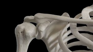
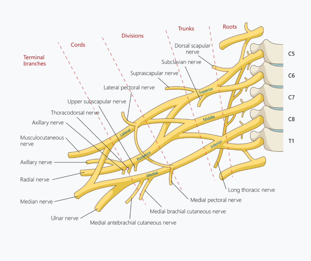
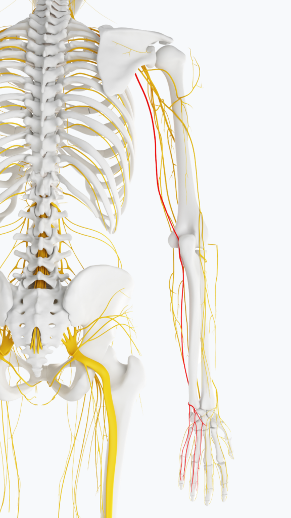
The ulnar nerve carries fibers primarily from the C8 and T1 spinal nerve roots and arises from the medial cord of the brachial plexus. It travels down the medial arm, pierces the medial intermuscular septum, and passes behind the medial epicondyle of the humerus. This superficial position creates the familiar electric sensation produced by striking the “funny bone.”
At the elbow, the nerve enters the cubital tunnel, a fibro-osseous channel between the medial epicondyle and olecranon. The elbow joint capsule and posterior portion of the ulnar collateral ligament contribute to the floor. The cubital tunnel retinaculum, often called Osborne’s ligament, forms the roof. Distally, the nerve passes beneath the aponeurosis connecting the humeral and ulnar heads of the flexor carpi ulnaris and continues into the forearm.
The ulnar nerve supplies the flexor carpi ulnaris and the ulnar portion of the flexor digitorum profundus. These muscles contribute to wrist flexion, ulnar deviation, and flexion of the distal joints of the ring and little fingers.
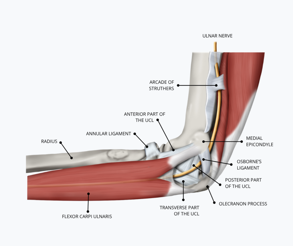
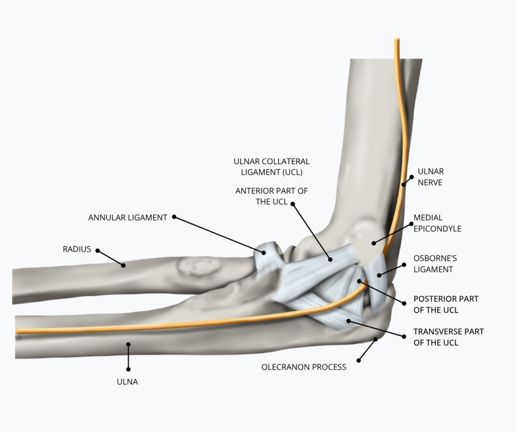
At the wrist, the nerve enters the hand through Guyon’s canal and supplies most of the intrinsic hand muscles. These include the interossei, hypothenar muscles, third and fourth lumbricals, and adductor pollicis. Together, they support grip, pinch, finger coordination, and precise hand movements. Sensory branches provide feeling to the little finger, the ulnar half of the ring finger, and the adjacent palm and dorsum of the hand.
For healthy function, a peripheral nerve must glide, deform, and maintain its internal blood supply as the joints around it move. Small blood vessels called the vasa nervorum supply oxygen and nutrients to the nerve fibers and their connective tissues. Sustained compression or excessive tension can restrict this circulation and impair nerve conduction.
Elbow flexion lengthens the ulnar nerve’s path around the medial epicondyle and reduces the space inside the cubital tunnel. The nerve normally accommodates these changes by moving within its connective tissue bed and distributing strain along its length. When the elbow straightens, tunnel dimensions and neural tension decrease.
Dysfunction and Its Causes
Cubital tunnel syndrome develops when compression, traction, or friction exceeds the ulnar nerve’s ability to adapt:
- Sustained elbow flexion narrows the tunnel and increases pressure within the nerve.
- Direct pressure against a table or desk (e.g., leaning on an elbow) adds compression where the nerve has little soft-tissue protection.
- Sleeping with the elbow tightly flexed.
- Repeated elbow flexion and extension may also irritate an unstable ulnar nerve that slips out of its groove and snaps over the medial epicondyle.
- Sports like baseball pitching, javelin throwing, and other activities that repeatedly load the inner elbow.
- Computer-based desk work, especially when the person rests the medial elbow on a desk or armrest.
- Professional driving, which may involve prolonged elbow flexion or resting the elbow against the door or window ledge.
- Prior fractures or dislocations, arthritic changes, osteophytes, joint swelling, ganglion cysts, and an accessory anconeus epitrochlearis muscle can reduce available space.
- Metabolic conditions such as diabetes may also increase a peripheral nerve’s vulnerability to compression.
The mechanical stresses described previously can impair intraneural circulation and disrupt axonal transport. Early in the process, reduced blood flow and localized damage to the nerve’s insulating myelin slow nerve conduction, producing intermittent tingling or numbness.
Continued compression may cause intraneural edema and greater sensitivity to movement. Severe or prolonged compression can damage axons, leading to persistent sensory loss, reduced grip and pinch strength, impaired finger coordination, and visible wasting of the intrinsic hand muscles.
Cautions
Symptoms in the ring and little fingers do not automatically confirm cubital tunnel syndrome. A C8 or T1 radiculopathy, lower brachial plexus disorder, ulnar nerve compression at Guyon’s canal, or generalized peripheral neuropathy may create a similar presentation.
Progressive weakness, persistent numbness, loss of dexterity, or visible hand-muscle wasting requires medical evaluation. Sudden symptoms following trauma, marked swelling, or rapidly worsening neurologic changes also warrant referral.
Assessment Considerations
A whole-system assessment considers the cervical and thoracic spine, shoulder girdle, elbow, forearm, wrist, and hand. Restricted shoulder motion may require the elbow and forearm to compensate during reaching. Limited elbow extension may keep the nerve under sustained tension, while hypertonicity in the medial arm and flexor-pronator region may reduce adaptability around the tunnel. The goal is to improve movement options, reduce unnecessary load, and create a safer mechanical environment for the nerve.
Preparing for Nerve Gliding
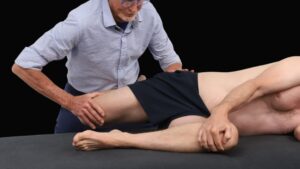
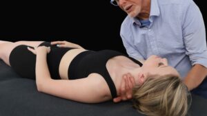
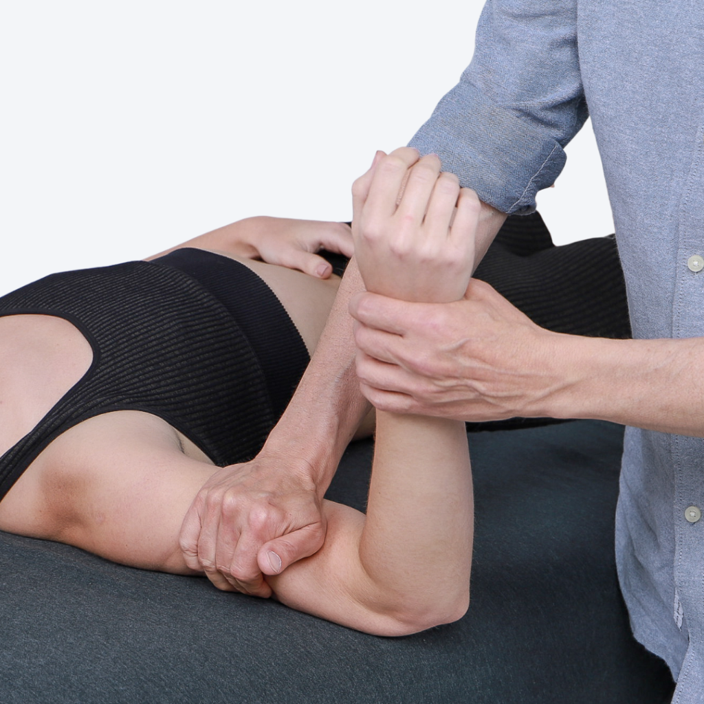
In the Treating Trapped Nerves course, Erik teaches a comprehensive soft-tissue sequence to prepare the ulnar nerve and its surrounding tissues for nerve flossing and tensioning. Rather than moving directly into neural mobilization, he works from proximal to distal, beginning with tissues that influence the C8-T1 nerve roots and lower trunk of the brachial plexus and continuing through the shoulder, medial arm, elbow, and forearm. The goal is to reduce protective muscle tone, improve cervical and shoulder-girdle mobility, and decrease unnecessary mechanical load before asking the ulnar nerve to move.
Focus on muscles that influence the position and movement of the cervical spine, clavicle, scapula, shoulder, elbow, and forearm, including:
- Anterior and middle scalenes
- Upper trapezius and levator scapulae
- Subclavius and pectoralis minor
- Latissimus dorsi and teres major
- Medial triceps
- Flexor carpi ulnaris and the surrounding flexor-pronator muscles
Warming and reducing excessive tone in these regions creates more movement options along the ulnar nerve pathway. Work around the medial elbow broadly and cautiously, avoiding direct pressure over the ulnar nerve and cubital tunnel. When the tissue is properly prepped, move into ulnar nerve gliding and tensioning.
Ulnar Nerve Set Up
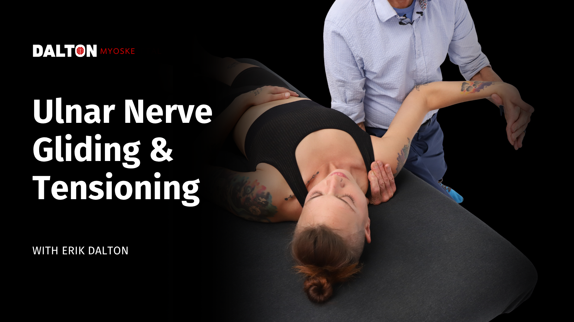
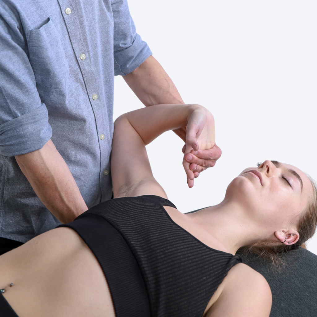
Stand on the right side of your client and grasp their right hand. With your left hand, lift the client’s arm out to the side to shoulder height and turn it outward from the shoulder, positioning the arm at 90 degrees.
Slide your right hand under the client’s shoulder and press down gently to stabilize the shoulder girdle. Bend the client’s elbow and brace it against your hip, continuing to bend it toward end range. This is what narrows the cubital tunnel and begins loading the nerve.
Turn the client’s forearm so the palm faces down. With your left hand, bend the wrist and fingers back and tip the hand toward the thumb side, paying particular attention to the fifth digit.
Bring the bent wrist toward the client’s head. Continue holding the fingers and wrist in this position while directing the client to bend and turn their head to the left, away from the arm you’re working, placing the nerve under increased tension. These movements should not increase pain.
Ulnar Nerve Gliding
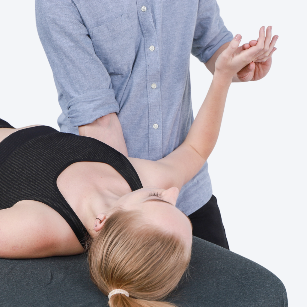
Keeping the elbow bent, bring the client’s wrist and fingers back to neutral and begin gliding the nerve. Ask the client to bend and turn their head to the left, then return to neutral, continuing this motion at a slow, rhythmic pace.
As the client’s head returns to neutral, bend the wrist and fingers back and tip the hand toward the thumb side, bringing it closer to the client’s head. Release the wrist back to neutral as the client bends and turns their head away. Continue this alternating pattern, tensioning one end while the other releases, for at least five gliding maneuvers.
Ulnar Nerve Tensioning
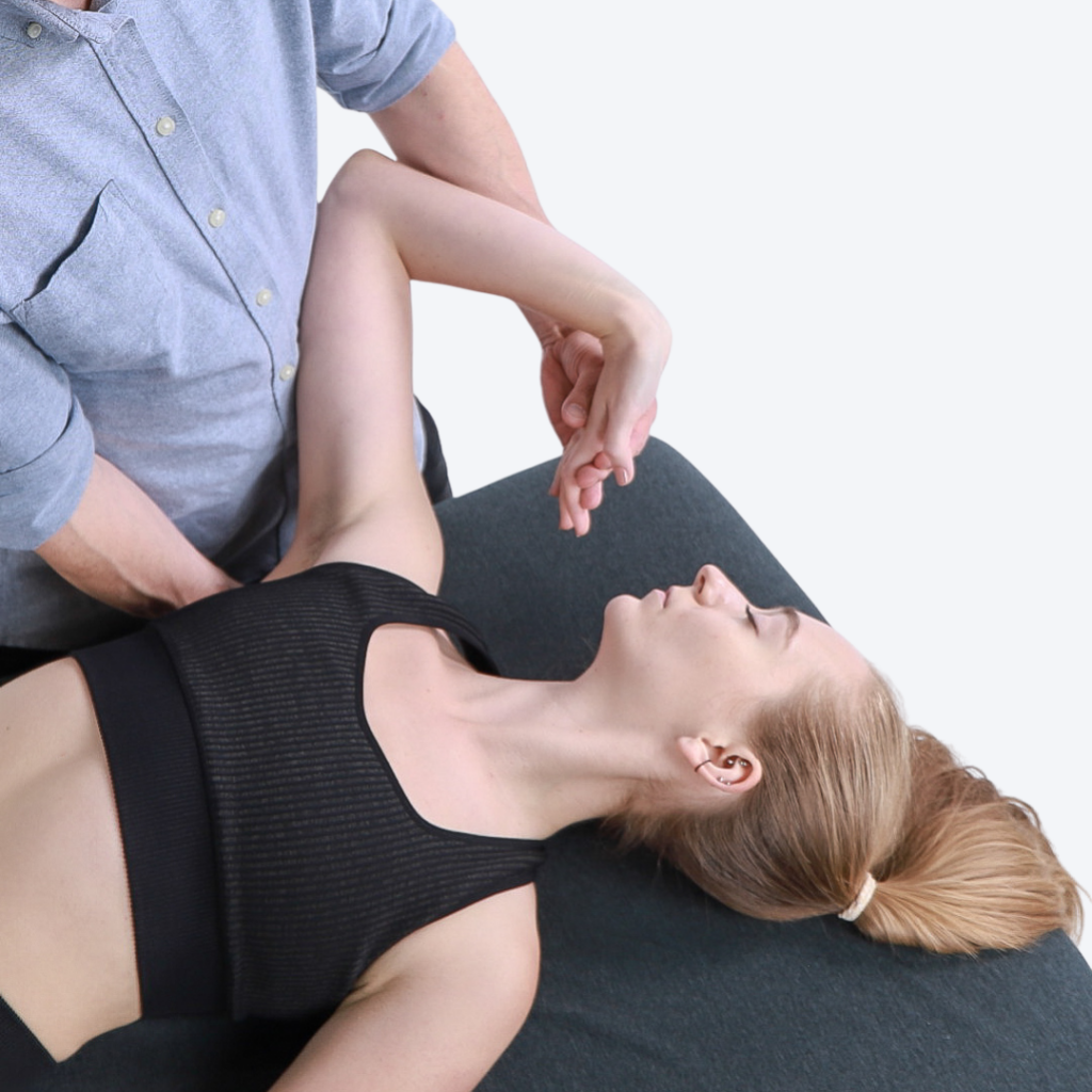
From the bent and turned head position, move into tensioning the ulnar nerve.
Keeping the elbow bent, bend the wrist and fingers back and tip the hand toward the thumb side as the client bends and turns their head to the left, bringing the hand closer to the client’s head to load the nerve at both ends at once. Hold to a count of five, release, and repeat three more times.
Want me to drop this into the Word document in place of the original three sections, either as a clean replacement or as tracked changes?
In Closing
Cubital tunnel syndrome reminds us that symptoms in the hand may reflect mechanical stress occurring anywhere along the ulnar nerve pathway. Effective treatment looks beyond the cubital tunnel to the cervical spine, shoulder girdle, arm, elbow, forearm, and wrist. By preparing these regions from proximal to distal, therapists create more movement options before introducing nerve gliding and tensioning. Neural mobilization offers therapists a practical strategy for reducing unnecessary neural load and helping clients move, work, and sleep with greater comfort.