Introduction
Knee pain is one of the most common complaints massage therapists encounter. Clients often arrive convinced their knee is “wearing out,” structurally damaged, or headed for surgery. And while degenerative changes do happen with age and use, research tells a surprising story: structural findings like meniscal tears, cartilage damage, and bone spurs show up frequently on imaging of pain-free people and correlate poorly with who actually has symptoms.1–3
This disconnect between what we see on an MRI and what someone feels points us toward a different explanation. Most knee pain isn’t caused by the knee failing. It’s caused by altered mechanics above and below the joint, combined with a sensitized nervous system that amplifies the problem.
Healthy Mechanics
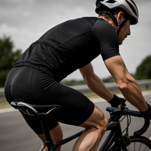
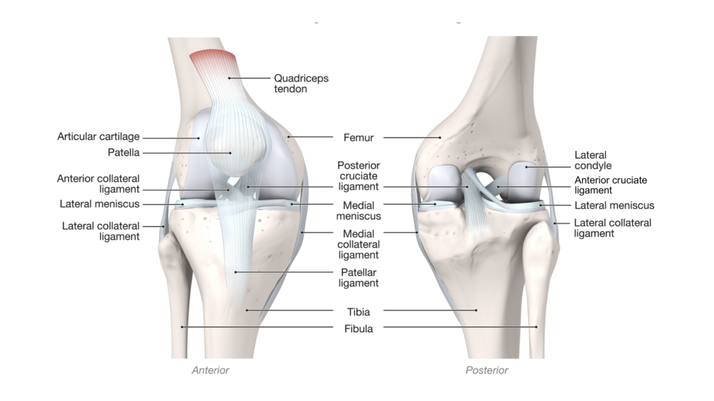
The knee serves as a transmission point in the lower extremity, transferring load between the foot and the pelvis during movement. The knee is described as a modified hinge joint because it allows slight rotation while handling compression forces, especially during the constant weight shifts of walking and running.
Healthy knee function depends on the coordinated interaction of several interdependent systems. The bony architecture of the tibiofemoral and patellofemoral joints provides the structural framework, while the menisci distribute compressive load, enhance joint congruency, and contribute to shock absorption. The cruciate and collateral ligaments guide and constrain movement within safe ranges, and the joint capsule provides both mechanical support and proprioceptive feedback. Surrounding this are the dynamic stabilizers, including the quadriceps, hamstrings, gastrocnemius, and popliteus, which actively modulate joint forces in response to changing load demands.
Central to healthy knee mechanics is accessory joint motion. These are the small gliding, rolling, and spinning movements within the joint that allow full range of motion without tissue strain. Equally important is myofascial force transmission, the way tension and load are distributed through fascial continuities rather than isolated muscles alone, meaning the knee is constantly responding to mechanical inputs from well beyond its immediate structures.
Dysfunctional Mechanics
When something disrupts neuromuscular coordination, joint mobility, or the smooth transfer of force through the lower extremity, whether from a restricted joint, muscle imbalance, or compensatory movement pattern, the knee ends up absorbing stress it was never designed to handle alone.
This is why treating knee pain without looking upstream and downstream usually produces short-lived results. Understanding how each region contributes to knee loading helps explain why a whole-system approach creates more lasting change.
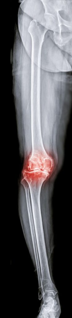
Foot and Ankle Restrictions: Limited ankle dorsiflexion is one of the most common contributors to knee dysfunction. When the ankle cannot move freely through its full range, the tibia cannot glide forward over the foot during the stance phase, forcing the knee to compensate with excessive rotation or early heel rise. Chronic ankle restrictions, whether from prior sprains, fascial thickening, or reduced subtalar mobility, alter ground reaction force transmission and change the timing and distribution of load arriving at the knee. Over time, these compensations disrupt neuromuscular coordination and increase compressive stress on the menisci, articular cartilage, and patellofemoral joint.
Loss of Tibial-Fibular Glide: The knee relies on intrinsic roll-glide mechanics to move smoothly. During knee flexion in a closed chain (like a squat), the femur must both roll backward and glide forward on the tibia to maintain proper joint congruency. It’s a combined motion, so if one part is missing, normal joint mechanics are disrupted.
The proximal tibiofibular joint allows the fibular head to glide superiorly/inferiorly and rotate slightly in response to tibial rotation and ankle dorsiflexion. When this joint becomes restricted, it can cause localized lateral knee pain that may be misattributed to iliotibial band friction. While the fibula serves as an important attachment site for lateral knee structures (LCL, biceps femoris), restrictions here don’t significantly alter knee joint mechanics or cruciate ligament tension.
Restricted accessory motion also sends threat signals to the nervous system, triggering protective muscle guarding in the hamstrings, peroneals, and lateral thigh. This guarding further limits movement and perpetuates pain issues.
Femoral Rotation and Patellar Tracking: Chronic postural habits, repetitive movement patterns, and compensations from old injuries can result in habitual hip rotation patterns where the femur rests in relative internal or external rotation. This rotational malalignment has profound effects on patellar tracking and tibiofemoral joint loading.
For example, when the femur remains internally rotated, the femoral trochlear groove (the channel on the front of the femur where the kneecap glides) rotates medially beneath the patella. This creates relative lateral displacement of the patella and increases compression along its lateral edge. Over time, this can contribute to anterior knee pain, grinding sensations (crepitus), and apprehension when going up stairs or squatting.
Restoring rotational freedom between the femur and its surrounding fascial envelope improves patellar tracking and reduces strain on surrounding tissues without forcing movement through the knee itself.
Patellofemoral Pain and Lateral Tracking: The patella, embedded within the quadriceps tendon, responds to forces imposed upon it from the thigh and lower leg. When lateral thigh tissues become hypertonic, particularly the vastus lateralis and iliotibial band, the patella is pulled laterally and compressed against the femoral groove.
Hip Flexor Tone and Anterior Knee Mechanics: Equally relevant is the relationship between hip flexor tone and anterior knee mechanics. When the rectus femoris remains chronically shortened, it pulls the patella upward, concentrating forces on areas of cartilage and limiting hip extension, causing compensatory stress at the knee. Restoring balance in these surrounding tissues allows the patella to track more centrally and the knee to move with less resistance.
MAT for Painful Knees
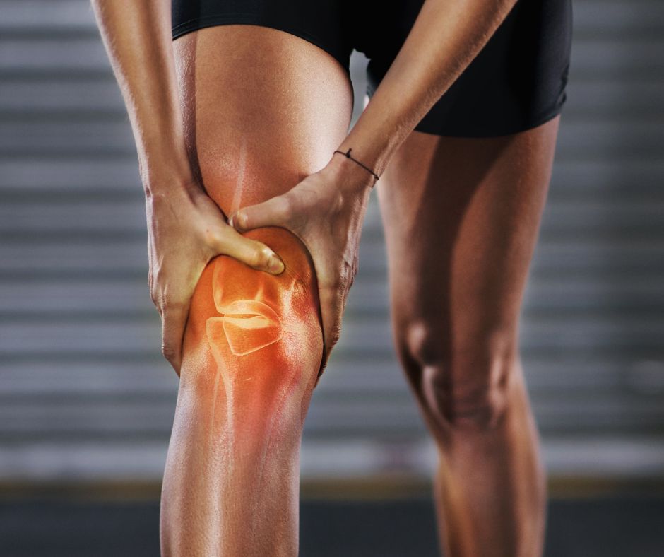
MAT takes a holistic approach to painful knees, addressing upstream and downstream influences on knee mechanics. The most productive areas to address include:
Below the knee: Restore ankle dorsiflexion mobility, address restrictions in the talocrural and subtalar joints, and release fascial tension through the posterior lower leg that may be limiting tibial advancement during gait.
At the knee: Re-establish accessory joint motion, particularly tibial-fibular glide and tibiofemoral roll-glide mechanics, and reduce protective guarding in surrounding muscular tissue. These small accessory movements are essential for pain-free range of motion.
Above the knee: Address femoral rotation restrictions within the fascial envelope of the thigh, and release chronic tension in the hip flexors (particularly rectus femoris) and lateral thigh structures that alter patellar tracking.
The three techniques that follow target proximal tibial-fibular glide, femoral rotation within its fascial sleeve, and knee joint accessory motion. Erik’s lower extremity knee routine is extensive, so the goal here is to try incorporating three effective MAT techniques into your current lower extremity routines to support session outcomes related to painful knees.
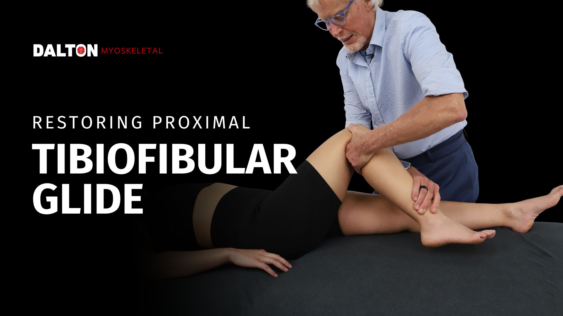
Restoring Proximal Tibiofibular Glide
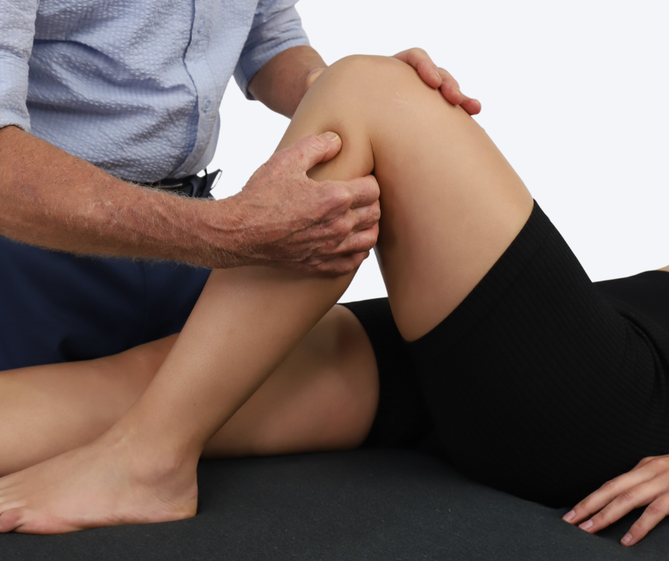
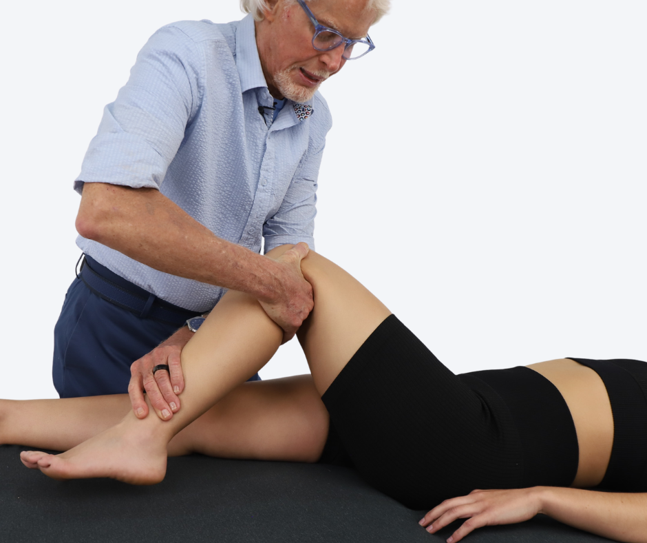
With the client supine, flex the knee and rest the client’s foot securely on the therapy table to stabilize the lower leg. With the fingers of your right hand, palpate the proximal fibular head and assess it for anterior and posterior glide. There should be a small amount of joint play.
Place your right hand around the proximal posterior calf and use the edge of your hand or knuckle as a wedge against the fibular head. Your left hand controls knee flexion by grasping the lower leg just above the ankle. Move the client’s knee into greater flexion with slight external rotation of the tibia. As the knee moves into flexion, your knuckle helps guide the fibular head back into proper alignment with the tibia. Move the client’s knee through flexion and extension as you continue to apply gentle posterior-to-anterior pressure on the fibular head. See the video to practice along with Erik.
The Log Roll Technique
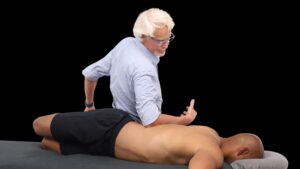
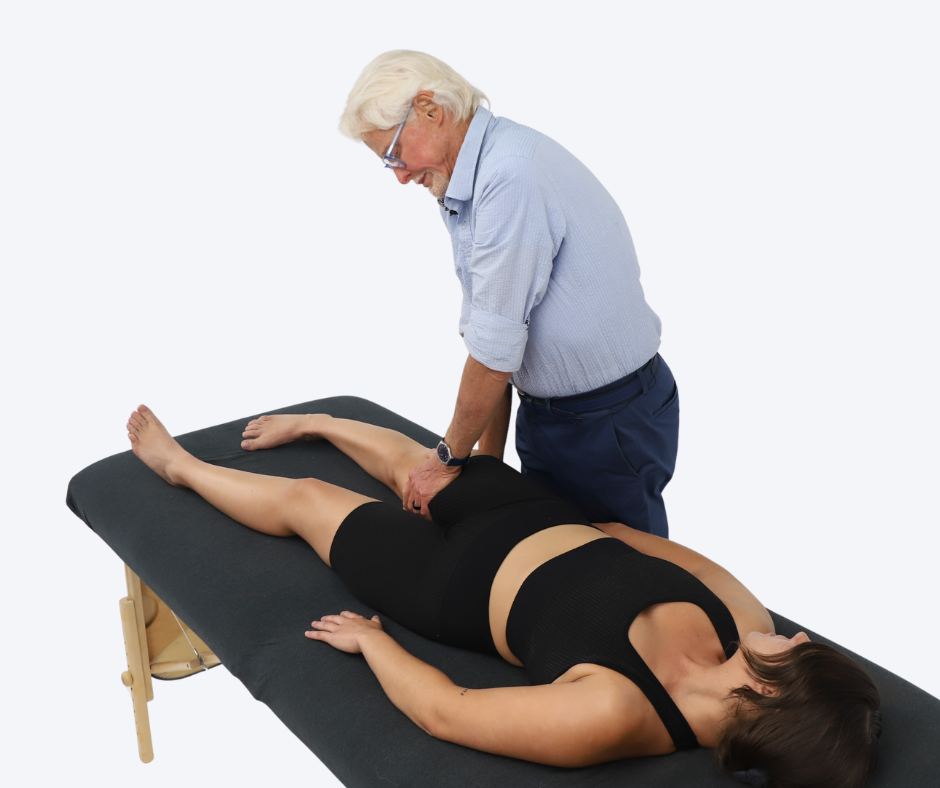
With the client supine and the knee extended, stand beside the table and snake your right hand under the client’s distal thigh while your left hand secures the anterior thigh fascia just above the knee. With both arms extended and your body providing the force, gently guide the client’s leg into internal rotation until you meet the first resistance barrier, maintaining knee extension throughout. Pin the thigh myofascia with your left hand and instruct the client to slowly begin externally rotating the entire leg against your resistance, visualizing the femur rolling within its fascial sleeve, using only as much effort as is comfortable for a slow count of five before fully relaxing. As the client relaxes, move the thigh into increased internal rotation to the new barrier, maintaining soft, continuous contact. Repeat this sequence several times, traveling superiorly and inferiorly along the thigh to restore rotational freedom. If the client presents with a fixed internal rotation pattern, reverse the maneuver. All movement should remain controlled, coordinated, and pain-free.
Menisci Technique
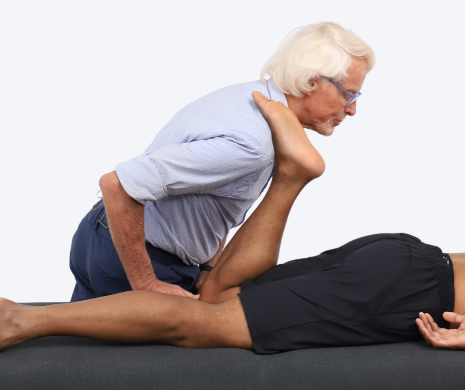
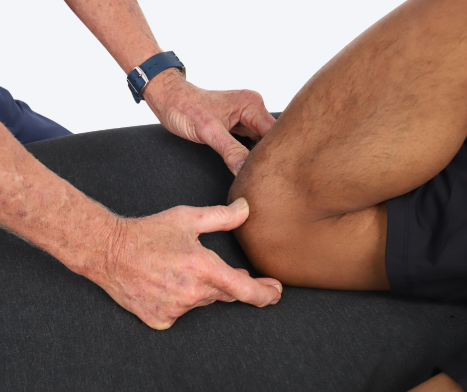
This technique improves accessory motion at the knee and ensures the menisci translate freely during movement. With the client prone, flex the client’s knee and place their foot on your shoulder so the lower leg is fully supported and controlled by your body rather than your hands. Place both thumbs into the soft tissue recesses at the medial and lateral joint lines, just inferior to the femoral condyles, where the joint capsule and meniscal horns are most accessible to palpation. This contact point sits slightly posterior to the patella.
Instruct the client to press the foot firmly into your shoulder for a slow count of five while you maintain steady thumb contact. This isometric contraction engages the posterior chain and creates a brief period of controlled joint loading. After five seconds, instruct the client to relax.
As the client relaxes, shift your body weight forward to gently guide the knee into greater flexion. Sustained thumb contact reduces periarticular tissue tension and supports normal joint mechanics as flexion increases, allowing the menisci to translate posteriorly as they would in healthy movement. Repeat the sequence two to three times.
In Closing
By recognizing the knee as a transmission point within the kinetic chain rather than a standalone joint, therapists can address the upstream and downstream restrictions that drive compensatory stress and pain. Restoring mobility at the ankle, freeing femoral rotation within its fascial envelope, and re-establishing accessory motion at the knee itself creates the mechanical foundation for healthier movement patterns.
References:
- Bianco Prevot L, Bensa A, Randelli PS, Filardo G. Cartilage lesions are not the main factor influencing pain and functional impairment in early knee osteoarthritis: a multivariate analysis on intra- and extra-articular factors affecting symptoms in over 200 knees. Bone Jt Open. 2025 Jul 16;6(7):828-835. doi: 10.1302/2633-1462.67.BJO-2025-0010.R1. PMID: 40665789; PMCID: PMC12264569.
- Englund M, Guermazi A, Gale D, Hunter DJ, Aliabadi P, Clancy M, Felson DT. Incidental meniscal findings on knee MRI in middle-aged and elderly persons. N Engl J Med. 2008 Sep 11;359(11):1108-1115. doi: 10.1056/NEJMoa0800777. PMID: 18784100; PMCID: PMC2897006.
- Sahota B, Howes M, Rio E, Docking S, Cook J. Association between knee pain location and abnormal imaging or arthroscopic findings: A systematic review. Ann Phys Rehabil Med. 2022 Apr 27;65(4):101638. doi: 10.1016/j.rehab.2022.101638. Epub 2022 Jan 13. PMID: 35093586; PMCID: PMC9650360.