Introduction
The lumbar spine is often asked to perform two demanding jobs at once. It must remain stable enough to support the upper body while also allowing us to bend, straighten, side-bend, and rotate during everyday movement.
Its five vertebrae, L1 through L5, have broad, weight-bearing bodies designed to manage substantial compressive loads. These vertebrae are larger and more robust than those in the cervical and thoracic regions and are separated by intervertebral discs that distribute compression, absorb shock, and permit controlled movement between adjacent segments.
The lumbar zygapophyseal joints, commonly called z-joints or facet joints, help guide spinal motion and protect against excessive rotation and translation. Their orientation favors flexion and extension while allowing smaller amounts of lateral flexion and rotation.
The Lumbosacral Junction
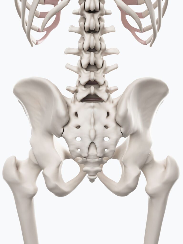
At the base of the lumbar spine, the fifth lumbar vertebra meets the sacrum at the lumbosacral junction (L5–S1). This transitional region must balance mobility with stability as it manages compression, shear, and the forces created during standing, bending, lifting, and walking.
This transitional zone transfers the weight of the trunk downward while receiving ground-reaction forces traveling upward through the kinetic chain. Because the sacrum slopes forward, body weight creates anterior shear, a force that encourages L5 to slide forward on S1. The disc, facet joints, ligaments, and surrounding muscles resist this shear while allowing controlled flexion and extension.
The lumbar spine, particularly the lumbosacral junction, must compensate for restricted hip extension or rotation, inadequate muscular control of pelvic position, altered gait, or limited thoracic mobility. Local dysfunction can also change how the region accepts and transfers load. Disc irritation, facet compression, segmental stiffness, instability, protective guarding, or spondylolisthesis may cause the lumbar spine to become the driver of compensation, forcing the hips, pelvis, or thoracic spine to alter their movements in response.
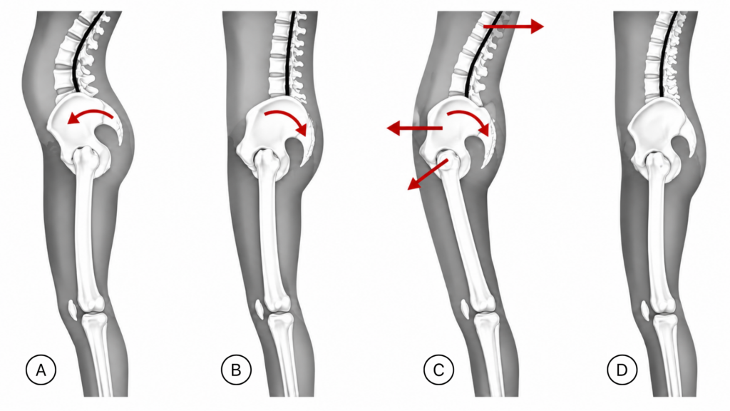
A. Anterior pelvic tilt causing a lordotic back. B. Posterior pelvic tilt causing a flat back. C. “Swayback” is caused when the upper trunk shifts backward to compensate for a forward-and-backward-tilted pelvis. The hip joints and knees hyperextend. D. Neutral pelvic and balanced posture.
The Lumbar Curve
The lumbar lordosis is part of the spine’s load-management system. Its shape reflects how the pelvis, hips, ribcage, abdominal wall, lumbar extensors, and nervous system organize the body against gravity and load. Pelvic position directly influences the curve.
When the pelvis tips anteriorly, the sacral base inclines forward and the lumbar spine moves toward greater lordosis and extension, increasing compression through the facet joints and lumbosacral junction (A above). Tight hip flexors and limited hip extension can reinforce this pattern by shifting more extension demand to the lumbar spine. The lumbar extensors may work harder, adding to fatigue and protective tension.
When the pelvis tips posteriorly, the sacral base moves backward, and the lumbar curve flattens, reducing available extension (B above). Tension through the hamstrings, gluteals, and posterior spinal tissues reinforce the tucked position. The lumbar spine may become less adaptable during standing, walking, or lifting, increasing stiffness and protective tension.
In a sway-back posture, the pelvis translates forward while the upper trunk shifts backward. The hips and knees often rest in hyperextension, and the lumbar curve appears flattened or lengthened. Because the body relies more on passive support from the joints, ligaments, and fascia, the nervous system increases lumbar extensor tone, contributing to stiffness, fatigue, and protective guarding.
Lumbar Compensation
Repeated compensation can make the lumbar region feel painful or unstable. The nervous system responds by increasing protective muscle activation. The deep stabilizers lose their coordinated, anticipatory timing, activating too late or inefficiently as movement begins. This reduces their ability to make the subtle adjustments that control motion between individual lumbar vertebrae.
Larger muscles take on more of the stabilizing work, while the thoracolumbar fascia becomes stiff and sensitive. Instead of distributing movement across individual segments, the lumbar spine begins moving as one protected block. Once this guarded pattern develops, reduced lumbar movement often creates compensation through the hips, pelvis, and thoracic spine.
MAT Techniques
Add the MAT techniques below to support improved movement, decrease protective muscle guarding, and reduce low-back pain.
Cautions
Recent lumbar surgery
Lumbar fusion
Spinal instability
Osteoporosis
Stenosis with neurological compromise
Acute low-back strain
Sharp or worsening pain
Numbness and weakness in the posterior legs
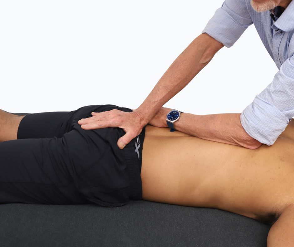
Sacral Base Decompression
This technique counteracts compressive forces by tractioning the sacrum inferiorly. Stand on the client’s left side towards the head of the therapy table. Place your right hand on the client’s sacrum and your left forearm on the thoracolumbar fascia.
Drop your body weight into the right hand with firm (not painful) pressure, moving the sacrum inferiorly. Your left forearm resists this motion at the thoracolumbar fascia by gently pulling the tissue headward. Ask the client to tilt their pelvis three or four times slowly to enhance this release.
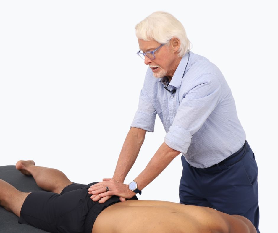
Sacral Roll
This technique mobilizes the sacrum to gently engage the sacroiliac ligaments.
Stand on the left side of the prone client and face their sacrum. Place your right hand on top of the client’s sacrum and your left hand on top of the right. Drop your body weight into both hands and begin rolling the sacrum counterclockwise.
Use your thenar eminence to move along the sacral border to contact the SIJ ligaments. Stay on the edge of the sacrum without dropping into the gluteal muscles as you move in a 360-degree circle around the bone.
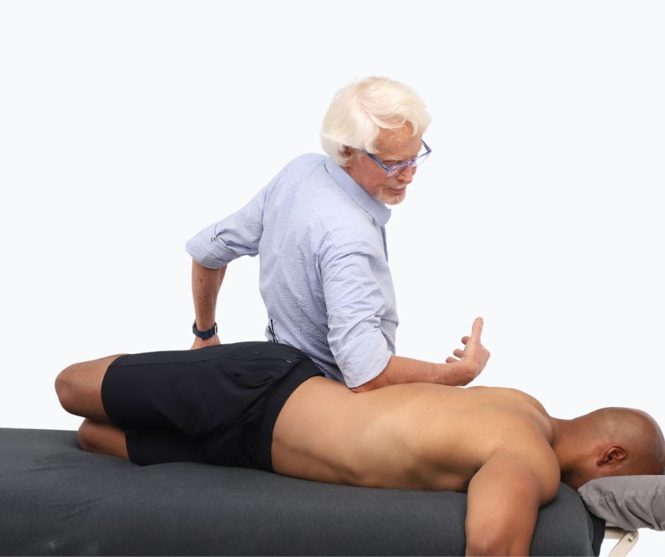
Modified Sims Counter-Rotation Technique
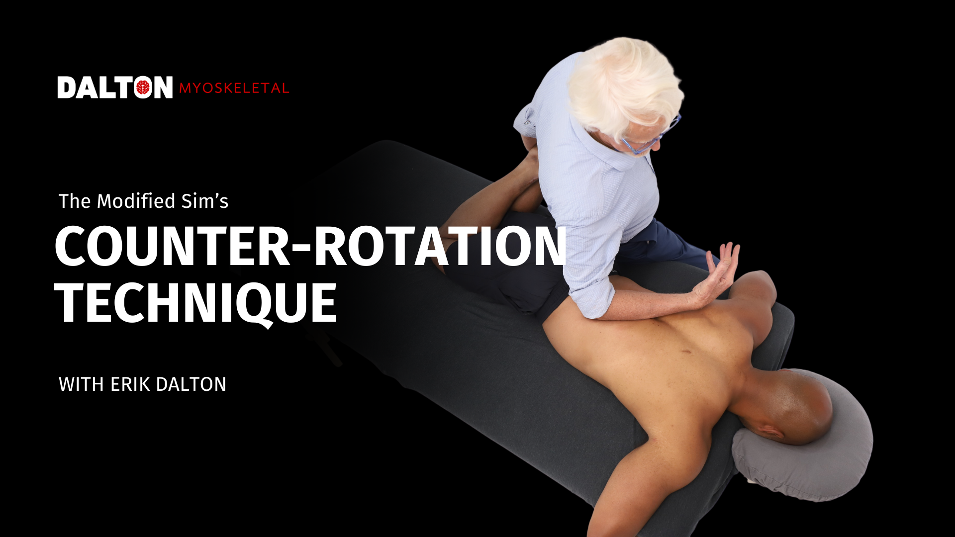
This technique is beneficial for clients with rotational restrictions or asymmetry through the lumbar and thoracic spine. Moving the flexed legs to one side uses the legs as an indirect lever to rotate the pelvis gently relative to the ribcage. The forearm applies an opposing force through the thoracolumbar tissues. This broad, controlled movement mobilizes multiple spinal segments without forcing rotation at any one joint. It also lengthens and loads the erector spinae, multifidi, and thoracolumbar fascia, encourages movement through the thoracic spine and ribcage, and provides novel sensory input that may help the nervous system reduce protective guarding.
The client is prone. Ask the client to bend both knees to 90 degrees and keep their knees close together. Stand on the client’s left side facing toward the client’s head, and hold the client’s ankles with your left hand. As you move the client legs to the left, the pelvis rotates to the right. Don’t force this rotational barrier. This position should feel comfortable to the client. Place your elbow and forearm on the right side of the client’s spine. Maintain the client’s leg position while slowly leaning your body weight into the forearm contact. The pressure should feel broad and comfortable, instead of sharp and deep. Sweep your forearm through the lumbar and thoracic paraspinals several times while continuing the counterforce twist using the client’s legs as a lever. Apply this technique on both sides of the body.
Elbow Low Back with Pelvic Tilts
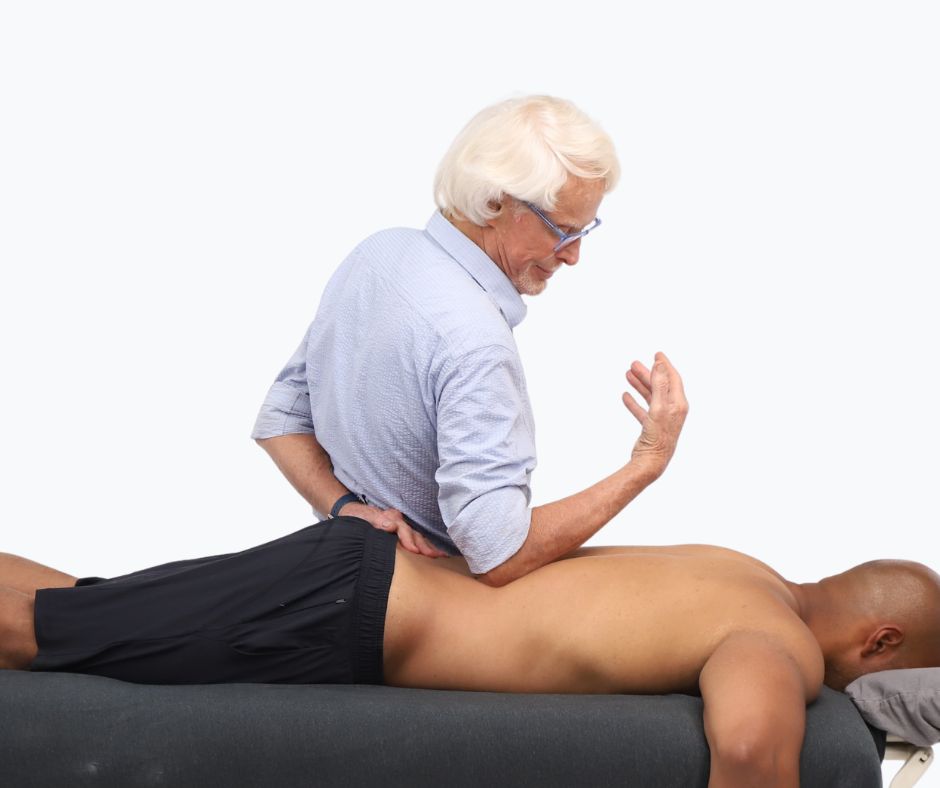
Active pelvic tilting adds movement to a sustained elbow contact. As the client alternates between anterior and posterior tilt, the lumbar tissues move beneath the therapist’s slowly gliding pressure. This movement-enhanced approach engages the erector spinae, quadratus lumborum, latissimus dorsi, and iliolumbar tissues while giving the brain novel sensory stimulation that helps reduce guarding.
Stand on the left side of the prone client. Make soft olecranon contact with the contralateral lumbar lamina groove. Sink slowly into the client’s first comfortable barrier using your body weight rather than shoulder tension and muscular effort. Ask the client to perform slow anterior and posterior pelvic tilts while you maintain steady contact or make slow gliding progress.
As the tissues soften, gradually follow the erector spinae laterally toward the quadratus lumborum, latissimus dorsi, and iliolumbar region. Move slowly and avoid compressing the twelfth rib, iliac crest, or lumbar spinous processes. Ask the client to stop tilting, slowly reduce your pressure and repeat the technique on the opposite side.
In Closing
The lumbar spine often becomes guarded when it is asked to manage more load or movement than it can comfortably distribute. These four techniques use decompression, mobilization, counter-rotation, and active movement to reduce protective tension and introduce safer movement options. Applied slowly and within a comfortable range, they can help the lumbar spine, pelvis, and thoracic region move and share load more efficiently.