Introduction
When clients come in because their low back feels tight, the front of their hips aches after a long day at the computer, they feel a pulling sensation through the groin, or they have trouble standing upright after sitting, the lower crossed model may help you make strong clinical decisions.
Consider a familiar case. A client works at a desk most of the day and drives another hour home. When she stands, her pelvis tips forward and her lumbar curve deepens. When walking, her leg does not extend far behind her body because the pelvis rolls forward instead, and the low back compensates to cover the missing hip extension. Palpation finds elevated tone in the lumbar erector spinae and tenderness through the anterior hip. When the client is prone and extends the hip, the pelvis lifts and the lumbar spine moves before the femur does.
Czech neurologist Vladimir Janda recognized that these findings often appear together and named the pattern lower crossed syndrome (also called pelvic crossed syndrome). This pattern offers us a useful clinical model for how muscle tone, recruitment, posture, and movement relate. Once we see the pattern, we can stop chasing isolated tender spots and ask which structures are carrying too much of the load, and which too little.
Understanding the Lower Cross
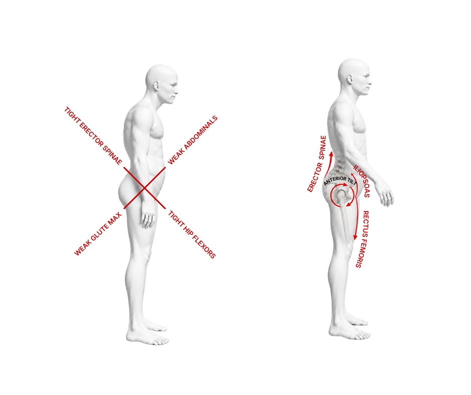
Viewed from the side, the muscular relationships Janda described show the hip flexors and lumbar extensors are shortened and hypertonic (iliopsoas, rectus femoris, tensor fasciae latae, and thoracolumbar erector spinae). The abdominal wall and gluteals, particularly gluteus maximus, are under-recruited, and poorly coordinated.
Calling the abdominals and gluteals weak can be misleading because they may test strong in isolation but still fail to contribute at the right time during standing, walking, lifting, or hip extension. With the pelvis tipped forward and the hip flexors resisting extension, the gluteals start from a poor position, and the nervous system recruits the lumbar extensors and hamstrings to finish the movement instead.
Why the Pattern Develops
Prolonged sitting is a common contributor because it holds the hips in flexion for hours and asks little from the gluteals. When the client stands, the nervous system falls back on the strategy it has practiced most, so the hip flexors stay guarded and the lumbar spine extends to keep the body upright.
Repetitive athletic positions, habitual standing posture, pain, pregnancy, stress, and poor movement coordination can produce similar relationships. A runner may repeatedly drive the hip flexors without developing adequate hip extension control. A weightlifter may brace by arching the low back instead of stacking the rib cage over the pelvis. A client protecting a painful hip may shorten the stride and recruit the lumbar extensors to avoid moving through the hip. The cause of the pattern differs, but the body arrives at a similar strategy.



MAT for Lower Crossed Syndrome
Myoskeletal Alignment Techniques (MAT) treats lower crossed syndrome as a coordinated lumbopelvic pattern, aiming to reduce lumbar extensor guarding, improve comfortable hip extension, and help the nervous system recruit the posterior hip muscles more effectively.
The four techniques that follow mirror the four elements of Janda’s cross. Elbow work paired with pelvic tilts addresses the hypertonic lumbar extensors. The anterior hip capsule technique improves extension through rectus femoris and the anterior capsular structures. The psoas stretch addresses the deeper hip-flexor component while the pelvis is stabilized. Spindle stimulation provides rapid sensory input to the under-recruited gluteals.
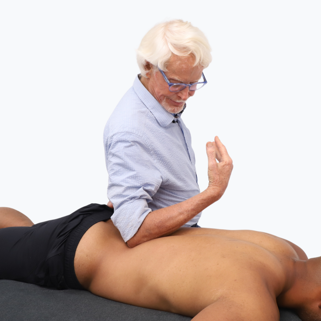
Elbow Low Back with Pelvic Tilts
The lumbar erector spinae form one of the hypertonic arms of the lower cross. In clients who habitually stand or move in lumbar extension, these muscles may remain active even during simple hip movement. The elbow contact provides broad compression and shear through the paraspinal tissues, while the client’s pelvic tilts add active movement beneath the contact
The client lies prone while you stand on their left side. Place a soft elbow in the contralateral lamina groove, lateral to the spinous processes. Sink gradually into the erector spinae and hook the tissue medial to lateral as the client performs slow pelvic tilts, letting the movement carry the tissue rather than forcing depth. Repeat three to five passes. Avoid direct pressure on bone and discontinue if the technique causes sharp or unfamiliar pain.
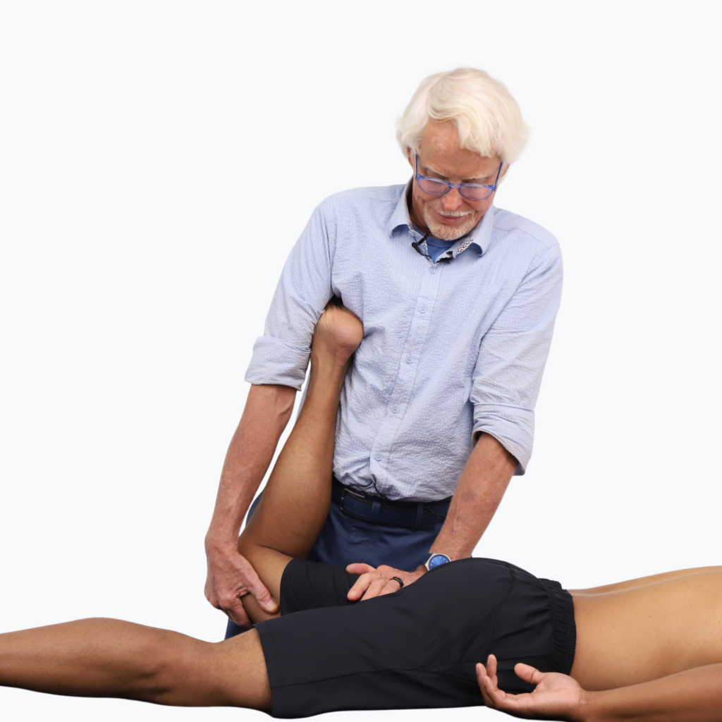
Anterior Hip Capsule Technique
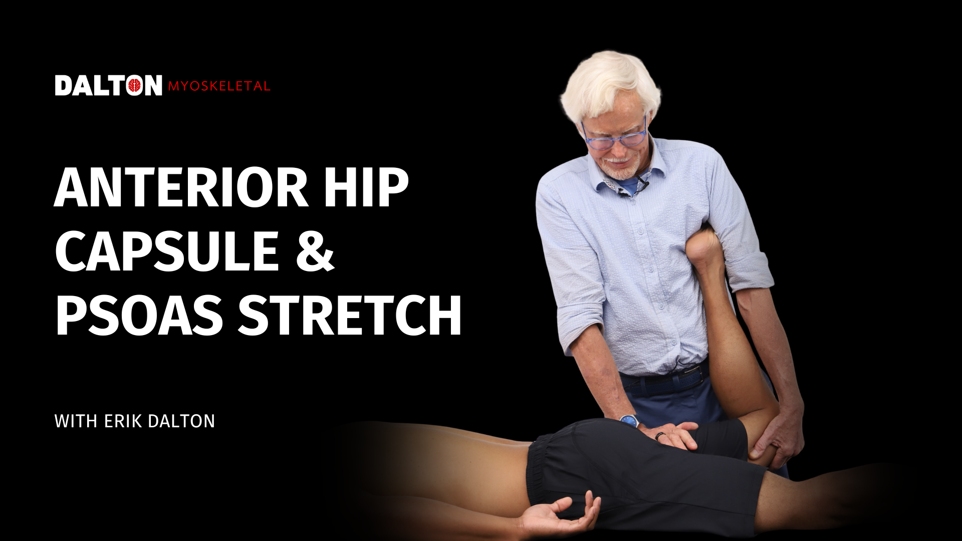
Lower crossed syndrome often includes an extension deficit at the hip. With the knee flexed, guiding the femur into extension lengthens rectus femoris and progressively loads the anterior capsule, letting the femur travel farther behind the pelvis during gait so the lumbar spine is less likely to compensate with more lordosis.
With the client prone, stand on their left side and flex the left knee to roughly 90 degrees, securing the foot beneath your right armpit and controlling the thigh with your right hand. Place your left palm on the posterior proximal femur just below the ischial tuberosity. As you step onto your left foot, guide the thigh into a small amount of hip extension while the left hand resists with a counterforce. Step rhythmically from foot to foot for up to two minutes, keeping the pelvis level and the front of the pelvis on the table. Stop for sharp anterior hip pinching, groin pain, lumbar compression, numbness, or tingling.
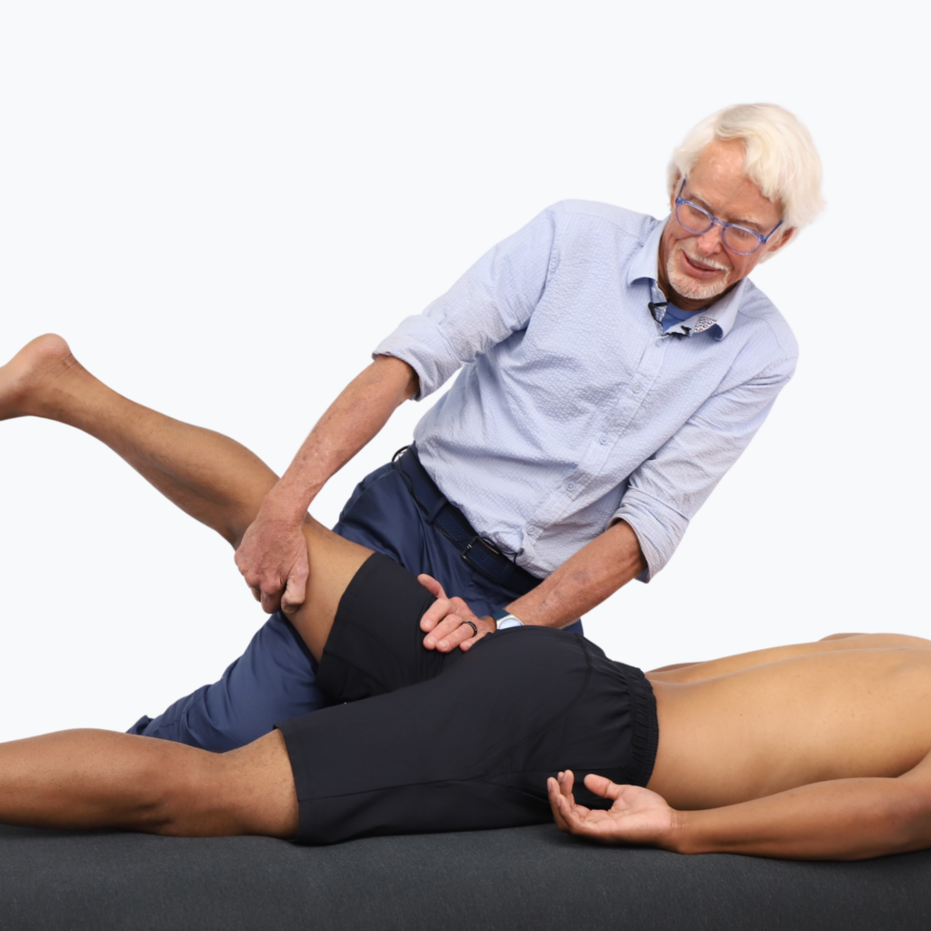
Psoas Stretch
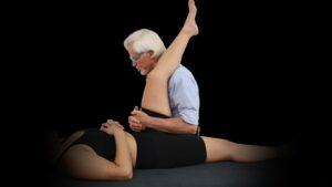
The iliopsoas connects the femur to the pelvis and lumbar spine and contributes strongly to hip flexion, making it a defining component of the lower cross. This technique uses hip extension with the knee straight, shifting emphasis away from rectus femoris and onto the iliopsoas. Stabilizing the posterior ilium is key. Otherwise, the pelvis tips forward, recreating the client’s habitual compensation instead of moving the hip.
The client remains prone while you stand on their left side. Place your left hand over the left PSIS to resist anterior pelvic tilt. Keep the client’s knee extended, support the distal thigh with your right hand, and lift the leg to the first comfortable hip-extension barrier, sliding your right knee beneath the anterior thigh for support. Ask the client to press the thigh gently downward into your knee for a count of five, then relax as you guide the femur into slightly greater extension with your hand. Repeat the contract-relax sequence three times. Groin pinching, increasing low-back pain, or neural symptoms are signals to stop.
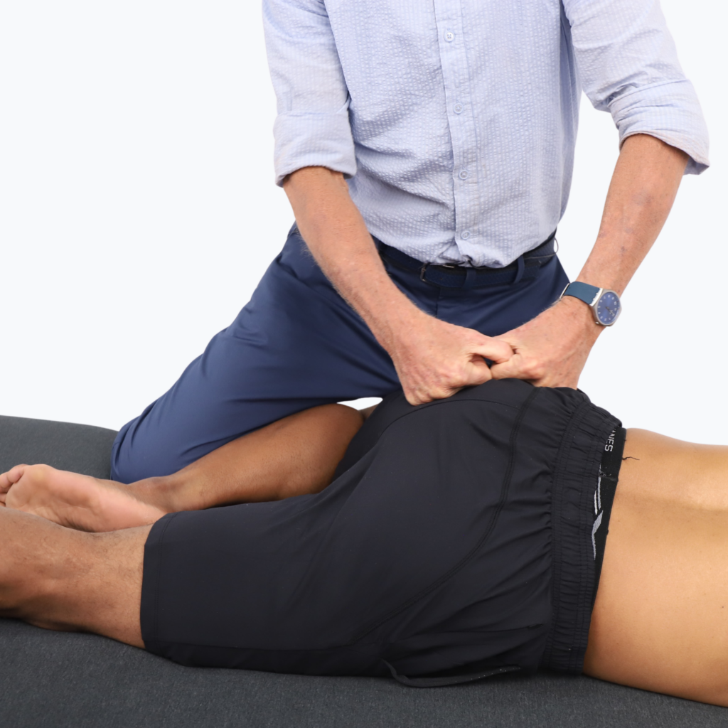
Spindle Stimulation of the Gluteals
Gluteus maximus should drive hip extension and help control the pelvis over the stance leg. When it is under-recruited, the hamstrings and lumbar extensors take over. Fast, rhythmic contact over the gluteal tissue provides dense proprioceptive input that improves recruitment of the posterior hip.
Place the client’s hip in a comfortable figure-four position to put the gluteal tissues on a mild stretch. Using soft fists, apply quick rubbing contacts across the gluteal muscles for one to two minutes. Avoid using tapotement. The movement should compress and move the tissue fibers in multiple directions.
In Closing
Lower crossed syndrome gives massage therapists a useful way to organize familiar findings: a tight low back, restricted anterior hip, anteriorly tilted pelvis, and poorly recruited gluteals are not isolated problems but parts of one movement strategy, in which the lumbar spine does more because the hips and trunk do less.
The four MAT techniques presented here address both sides of the cross: reducing tone in the lumbar extensors, improving anterior hip mobility, encouraging true hip extension, and helping the client rediscover the gluteals. When the hip can extend and the posterior chain contributes on time, the low back no longer has to create every movement or absorb every force alone.
Works Consulted
Jeon, I.-C., Hwang, U.-J., Jung, S.-H., & Kwon, O.-Y. (2016). Comparison of gluteus maximus and hamstring electromyographic activity and lumbopelvic motion during three different prone hip extension exercises in healthy volunteers. Physical Therapy in Sport, 22, 35–40. https://doi.org/10.1016/j.ptsp.2016.03.004
Keter, D. L., Bialosky, J. E., Brochetti, K., Courtney, C. A., Funabashi, M., Karas, S., Learman, K., & Cook, C. E. (2025). The mechanisms of manual therapy: A living review of systematic, narrative, and scoping reviews. PLOS ONE, 20(3), e0319586. https://doi.org/10.1371/journal.pone.0319586
Neumann, D. A. (2024). Neumann’s kinesiology of the musculoskeletal system: Foundations for rehabilitation (4th ed.). Elsevier.
Page, P., Frank, C. C., & Lardner, R. (2010). Assessment and treatment of muscle imbalance: The Janda approach. Human Kinetics.