fbpx

Low Back Z-joint Pain: Addressing Facet Joint Syndrome

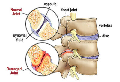
Clients with nonspecific low back pain may be experiencing mechanical wear and tear of the zygapophyseal joints, located in the posterior aspect of the lumbar spine (Image 1.). These bony articulations are commonly referred to as facet joints, but the proper terminology is zygapophyseal joints or Z-joints — derived from the Greek words zygos, meaning bridge, and physis, meaning outgrowth. The Z-joints are lined with cartilage, lubricated in synovial fluid, and covered by a tough joint capsule. When healthy, they allow smooth gliding and sliding while preventing excessive lumbar rotation. However, in our middle-aged and elderly population, normal vertebral motion is often compromised.

An injury, repetitive movements, obesity, weak posture, and aging articular cartilage may change the way the Z-joints align and move on one another. Trauma combined with prolonged gravitational exposure causes thinning of the joint capsule, cartilage degradation, and accompanying bone spurs. Similar to knee arthritis, these changes make it difficult for the joint to move fluidly, which often triggers an inflammatory response. With chemically sensitized medial branch sensory nerves bombarding the spinal cord and brain with danger signals, the back stiffens and may hurt during certain movements.

Acute episodes of lumbar Z-joint pain are typically intermittent, generally unpredictable, and occur a few times per month or per year. Although standing may be somewhat limited, sitting seems to be the most provocative position. In acute cases, Z-joint symptoms typically localize to one side of the back adjacent to the spine. In chronic cases, diffuse pain may spread into the buttock, groin, or down the entire limb.

Image 1. Z-joint Pathology: Mechanical wear and tear of the zygapophyseal (Z-joints)

Z-joint Pain Provocation Tests

In Image 2., I begin the assessment by gently palpating the paravertebral tissues overlying the lumbar transverse processes.
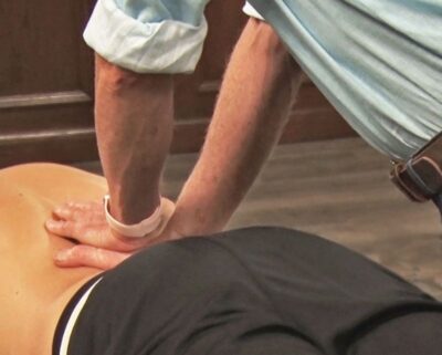
Image 2. Z-joint Pain Provocation Test: Using soft palms, the therapist palpates the paravertebral tissues overlying the lumbar transverse processes and the client reports Z-joint tenderness.

The Z-joints themselves are not manually palpable, but this maneuver is helpful in localizing and reproducing any point tenderness, which commonly accompanies Z-joint mediated pain.  If the client reports localized unilateral tenderness, there are several confirmation exams I’ve found helpful, especially the Sphinx Hyperextension Test and Kemp’s Test (Images 3 and 4).
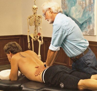
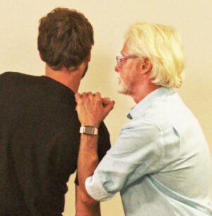
Image 3. Sphinx Hyperextension Test: The client assumes a pain-free sphinx position and the therapist's soft palms apply very gentle pressure to each side of the lumbar spine. Record as positive for possible Z-joint pathology if the client reports localized low-back or gluteal pain.
Image 4. Kemp's Test: The therapist asks the client to right rotate, right sidebend, and extend his torso. The therapist places both hands on the client's shoulder and gently depresses. Record as a positive if this maneuver reproduces or enhances the client's symptoms. However, if the pain follows a dermatomal pathway into the lower extremity, it may indicate sciatic nerve root compression.

In the Kemp’s Test, backward bending, rotation, and sidebending toward the affected side may elicit Z-joint symptoms. However, it’s important to note that this pain provocation test may also implicate nerve root compression and sciatica symptoms. Sharp buttock and leg pain following a dermatomal pattern may suggest sciatica, whereas Z-joint irritation is suspected if the test reproduces the client’s localized low back pain. If unsure, several other tests, such as the Slump Test and Straight Leg Raise Test, should be clustered to confirm your findings.

Restoring Z-joint Mobility

Numerous studies have examined conservative care for people with low back pain, but I’m unaware of any published investigations that have targeted Z-joint pain specifically. Nevertheless, most experts agree that the general principles of conservative treatment for nonspecific low back pain can be applied to Z-joint pain, too.1

I’ve had reasonable success treating both acute and chronic cases of Z-joint pain using the Myoskeletal Alignment Techniques (MAT) demonstrated in Images 5 and 6. These graded exposure stretching maneuvers are designed to bring balance to musculofascial tissues that torsion the pelvic bowl and create excessive anterior pelvic tilt. When performing these dynamic postural stretching routines, it’s best to address all connective tissues that articulate with the lumbar spine, hips, and legs and may be creating abnormal compressive loading through arthritic Z-joints.
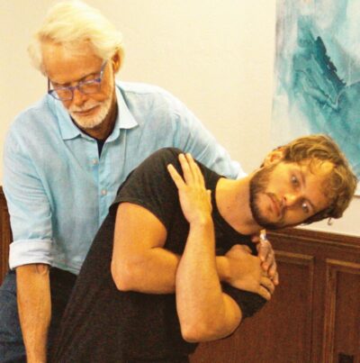
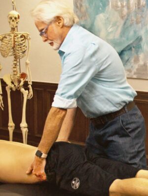
Image 5. Passive Sidebending Technique: With his right hand bracing the client's thigh, the therapist's left hand sidebends the client's lumbar spine to the left to stretch the quadratus lumborum and decompress the Z-joints on the client's right side. The client is asked to gently right sidebend against the therapist's resistance to a count of five and relax. The therapist once again left sidebends the client to the next restrictive barrier. Repeat 3-5 times and retest using the Kemp's test.
Image 6. Iliosacral Alignment Technique: The therapist's left hand lifts the client's left anteriorly/inferiorly rotated ilium and his right palm braces the right posterior superior iliac spine. The therapist gently pulls with his left hand while resisting with his right. The client is asked to gently push his left ilium towad the table to a count of five and relax. The therapist rotates the client's pelvis to the next restrictive barrier to restore Z-joint alignment.

In short, MAT treatment goals for Z-joint pain include:

  • educating the client by explaining their pain in a reassuring manner that avoids catastrophizing.
  • reducing excessive lumbar lordosis.
  • restoring lumbar spine mobility, pelvic alignment, and pain-free movement.
  • providing (playful) self-care advice to strengthen and stabilize the lumbopelvis.

Summary

During each therapy session, feel free to offer advice about various sitting and standing postures that may help relieve compressive stress through the Z-joints. Cueing the client about faulty movement patterns helps bring the brain’s attention to protectively guarded areas. All our movements are governed by the central nervous system, so the brain will limit flexibility, range of motion, and mobility if it feels there is a potential danger. Therefore, it’s important to always maintain good communication with our clients and keep them actively engaged in the therapy process. While Z-joint arthritis can’t be dramatically reversed, I’ve found that exercise, lifestyle changes, and proper bodywork can contribute to a better quality of life and less discomfort.

References

1. Binder, D., & Nampiaparampil, D. (2009). The provocative lumbar facet joint. Current Reviews in Musculoskeletal Medicine, 2(1), 15-24.

On sale this week only!

Save 25% off the Upper Body Course! 

NEW! Enhanced video USB format!!!

Learn unique approaches to work with the shoulder girdle, arms, neck, and torso, this course will prepare you to relieve painful myoskeletal issues in the upper body. Through video demos and animation, you’ll learn to identify several compensatory movement patterns and their associated reflexogenic pain. With this understanding of where the true source of problems arise, you’ll be able to develop highly effective treatment protocols and deliver lasting results. This course provides you with the skills you need to confidently relieve pain issues in the upper body. (20 CE)

Bonus: When you order the Home Study version, get the eLearning course and and an additional 2CE Ethics eCourse for free!

Save 25% off the Upper Body Course this week only. Offer expires Mon Sept 16th.  Click the button below for more information and to purchase the course for CE hours and a certificate of completion to display in your office. 

Free subscription to the Technique Tuesday Newsletter

Receive an in-depth article like the one on this page along with a technique video every week in your inbox with no subscription fees.