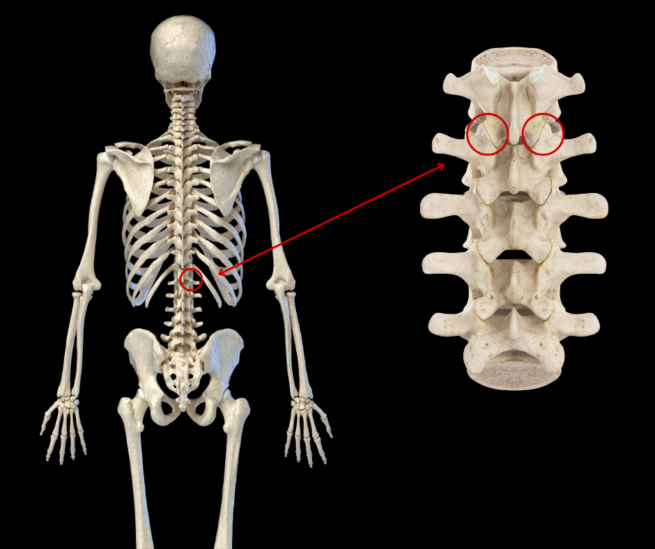
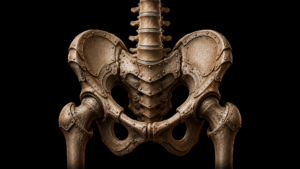
The zygapophyseal joints are a crucial component of the human vertebral column, contributing to its stability, flexibility, and load-bearing capabilities. Z joints are paired synovial joints formed from the superior articular processes of one vertebra and the inferior articular processes of the vertebra above. Z joints are the only synovial joints in the spine, with hyaline cartilage overlaying subchondral bone, a synovial membrane, and a joint capsule. Synovial fluid lubricates the joint, nourishes the articular cartilage, and facilitates smooth gliding of the articular surfaces.
Z joints function to stabilize the spine and limit axial rotation. Researchers estimate that Z joints carry approximately 25% of the dynamic axial load. Together with the intervertebral discs, Z joints help distribute the weight of the upper body evenly along the vertebral column preventing excessive stress on any single vertebra.
Zygapophyseal (Z) joints are paired synovial joints formed from the superior articular processes of one vertebra and the inferior articular processes of the vertebra above.
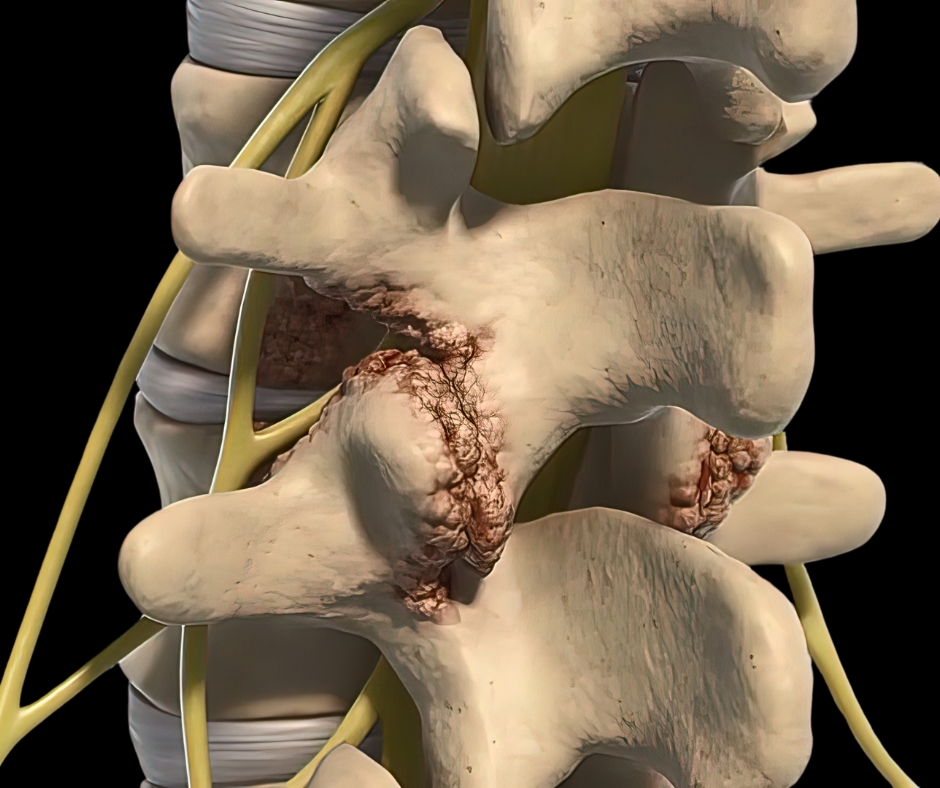
Like other synovial joints, Z joints can undergo degenerative changes over time in response to injury, trauma, obesity, age, and poor posture. The breakdown of the articular cartilage, development of bone spurs (osteophytes), and a reduction in the joint’s ability to absorb shock characterize osteoarthritis of the Z joints. As osteophytes form, the boney outgrowths place pressure on adjacent structures such as spinal nerves, intervertebral discs, ligaments, the facet joint capsule, and nearby muscles. The synovium often becomes inflamed, exacerbating pain and stiffness. Associated conditions, like intervertebral disc degeneration, narrow the disc space altering mechanical load distribution and resulting in progressive stress on Z joint articulations and joint capsules.
The musculoskeletal system responds to Z joint dysfunction by sending muscles like the multifidus, rotatores, interspinales, intertransversarii, erector spinae, and quadratus lumborum into protective spasm. These muscles attempt to stabilize the vertebral column causing additional pain, reduced movement, and inhibition of muscle function.
Z joints can undergo degenerative changes over time in response to injury, trauma, obesity, age, and poor posture. This image shows Z joint osteoarthritis.
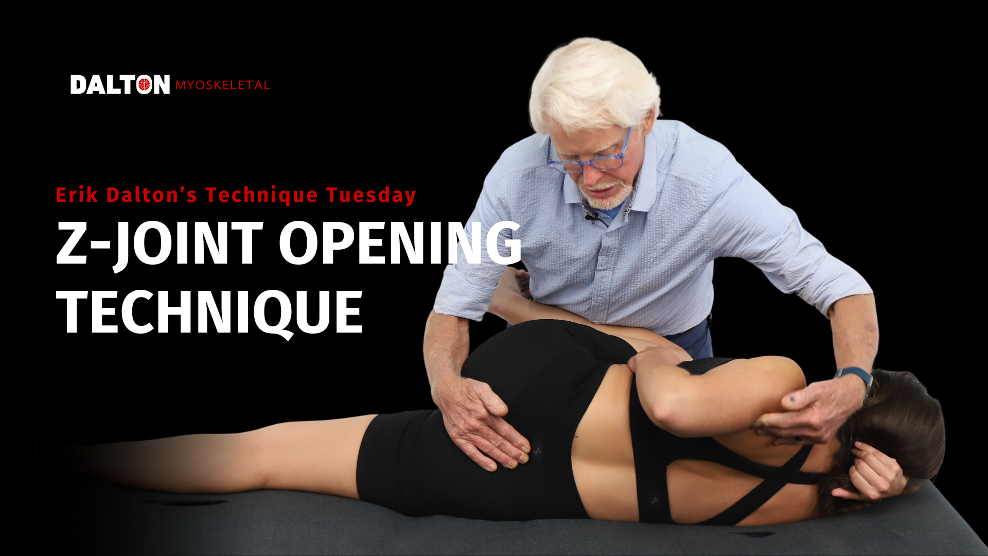
MAT for Z Joint Pain
Massage therapists are often quick to address low back issues with deep work on low back muscles, the rectus femoris, and iliopsoas. Master Myoskeletal Therapists® typically choose movement first, often with graded-exposure stretches that help reduce muscle tension and balance the innominates. As we expose the nervous system to new movement possibilities, we reduce the threat of pain. Soon, the body feels safe to down-regulate nervous system activity and drop protective muscle guarding. We can use deep friction, if necessary, but we are just as likely to apply muscle spindle activation techniques to wake up inhibited muscles and nerve flossing to free trapped nerves. Here, I’ll share two of the many methods we might choose that help improve Z joint function. You’ll find these methods are valuable additions to your bodywork toolbox.
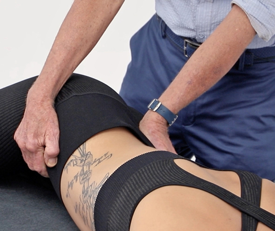
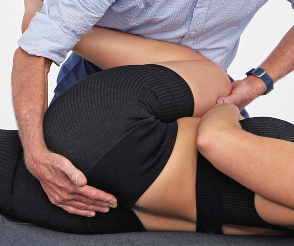
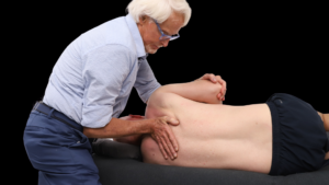
Iliosacral Alignment Technique (Above)
This is an indirect approach that helps level the sacral base, creating the conditions for proper Z joint alignment. With the client prone, your right hand lifts the client’s right ilium while your left palm braces the left posterior superior iliac spine. Gently pull with your right hand while resisting with your left. Ask the client to gently push their right ilium toward the table to a count of five and relax. You rotate the client’s pelvis to the next restrictive barrier to help restore Z joint alignment. Repeat this maneuver three to five times and then repeat it on the other side.
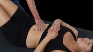
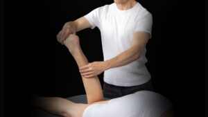
Z-Joint Opening (Above)
This technique decompresses the Z joints. Face the client and place the client’s knees in a slightly flexed position. Lift the client’s left flexed knee and place it against your belly. Your left hip supports the client’s leg so that when you step to your left foot, you initiate greater hip flexion. Contact the left L5 transverse process (or the lamina groove) with the fingers of your right hand. Now, step onto your left foot, flexing the client’s hip while you use the fingers of your right hand to drag the sacrum away from L5. Make sure your fingers are hooking deep in the groove and the client’s knee is flexed tight as you step onto your left foot. Move your fingers to L4 and repeat this process. Repeat this process with the client lying on the other side.
In Closing
In conclusion, low back pain is a prevalent and debilitating condition affecting a significant portion of the adult population. The zygapophyseal joints, or Z joints, are increasingly recognized as primary pain generators in low back conditions. While many techniques might be used to address Z joint pain, the Iliosacral Alignment Technique and Z-Joint Opening shown here give good results.