fbpx

Low Back Z-joint Pain: Addressing Facet Joint Syndrome

Understanding Z-Joint Pain: The Hidden Culprit Behind Nonspecific Low Back Pain

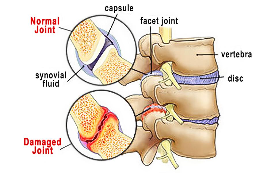
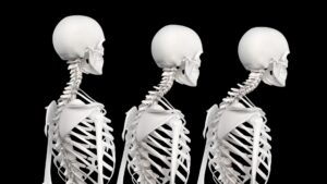
Many clients who come in with “nonspecific” low back pain may actually be experiencing mechanical stress in the zygapophyseal joints, also known as facet joints or Z-joints—small, paired articulations on the back of the vertebrae that guide spinal motion and share load with the intervertebral discs.

These joints are lined with cartilage, enclosed in a synovial capsule, and lubricated with fluid to allow smooth gliding between vertebrae. When healthy, Z-joints permit controlled motion, especially flexion, extension, and sidebending, while preventing excessive rotation that could damage the discs.

Unfortunately, wear and tear over time can compromise this elegant system.

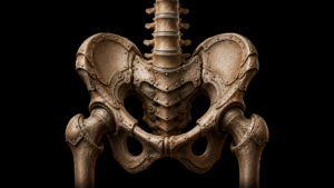
Anatomy of the Z-Joints

Each lumbar vertebra connects with the one above and below through two Z-joints. Together with the intervertebral disc, they form a three-joint complex that balances mobility and stability.

  • Structure: The articular surfaces are covered with hyaline cartilage and enclosed by a capsule rich in sensory nerves.
  • Function: They bear roughly 20–25% of compressive load during extension and up to 50% during rotation (Adams & Dolan, 2005).
  • Innervation: The medial branches of the dorsal rami transmit sensory information, explaining why inflammation here can cause sharp, localized pain near the spine.

Over time, repetitive strain, poor posture, injury, or degenerative changes can alter how these joints align and move—leading to inflammation, fibrosis, and bone spur formation.

How Z-Joint Dysfunction Develops

When cartilage thins and the capsule becomes lax or inflamed, motion becomes restricted and uneven. The resulting micro-instability leads to:

  • Increased friction between articular surfaces
  • Joint capsule irritation
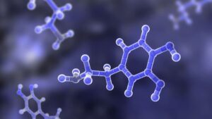
  • Chemical sensitization of the medial branch nerves

This chemical irritation can bombard the spinal cord with nociceptive signals, triggering muscle guarding and stiffness. In other words, the nervous system starts to “lock down” movement in an effort to protect the spine, ironically perpetuating the pain cycle.

Common contributors include:

  • Repetitive bending or twisting
  • Prolonged sitting or hyperlordosis
  • Obesity or deconditioning
  • Postural asymmetries or pelvic torsion
Image 1. Z-joint Pathology: Mechanical wear and tear of the zygapophyseal (Z-joints)
Z-joint Pathology: Mechanical wear and tear of the zygapophyseal (Z-joints)

Recognizing Z-Joint Pain Patterns

Z-joint pain typically presents as localized, unilateral low back pain that may occasionally radiate into the buttock or thigh but rarely below the knee.

  • Acute pain often appears intermittently and is triggered by extension or rotation.
  • Chronic cases may show more diffuse discomfort with morning stiffness or pain after prolonged sitting.

Clients often report that sitting or driving aggravates symptoms more than standing or walking.

Z-joint Pain Provocation Tests

Although Z-joints are not directly palpable, tenderness in the paravertebral tissues overlying the transverse processes may indicate Z-joint involvement.

Image 2. Z-joint Pain Provocation Test: Using soft palms, the therapist palpates the paravertebral tissues overlying the lumbar transverse processes and the client reports Z-joint tenderness.
Z-joint Pain Provocation Test: Using soft palms, the therapist palpates the paravertebral tissues overlying the lumbar transverse processes and the client reports Z-joint tenderness. Localized, unilateral tenderness suggests Z-joint irritation.

The Z-joints themselves are not manually palpable, but this maneuver is helpful in localizing and reproducing any point tenderness, which commonly accompanies Z-joint mediated pain.  If the client reports localized unilateral tenderness, there are several confirmation exams I’ve found helpful, especially the Sphinx Hyperextension Test and Kemp’s Test

Image 3. Sphinx Hyperextension Test: The client assumes a pain-free sphinx position and the therapist's soft palms apply very gentle pressure to each side of the lumbar spine. Record as positive for possible Z-joint pathology if the client reports localized low-back or gluteal pain.
Sphinx Hyperextension Test: Client lies prone in a comfortable sphinx position. The therapist applies gentle bilateral pressure beside the spine. Reproduction of localized low back or gluteal pain suggests Z-joint pathology.
Image 4. Kemp's Test: The therapist asks the client to right rotate, right sidebend, and extend his torso. The therapist places both hands on the client's shoulder and gently depresses. Record as a positive if this maneuver reproduces or enhances the client's symptoms. However, if the pain follows a dermatomal pathway into the lower extremity, it may indicate sciatic nerve root compression.
Kemp's Test: Client rotates, sidebends, and extends toward the affected side while the therapist places both hands on the client's shoulder and gently depresses.

With Kemp’s test, reproduction of localized back pain supports Z-joint involvement, while pain radiating down the leg suggests nerve root compression.

If uncertainty remains, use the Slump and Straight Leg Raise tests to differentiate discogenic or radicular pain.

Myoskeletal Approach: Restoring Mobility and Balance

While there are few studies isolating Z-joint pain specifically, research on conservative care for nonspecific low back pain applies here as well. Manual therapy, exercise, and movement retraining can significantly reduce symptoms and improve function.

Myoskeletal Alignment Techniques (MAT) address these dysfunctions by decompressing the affected joints, rebalancing muscle tone, and restoring symmetrical movement between the spine and pelvis.

The graded exposure stretching maneuvers illustrated below are designed to bring balance to musculofascial tissues that torsion the pelvic bowl and create excessive anterior pelvic tilt. When performing these dynamic postural stretching routines, it’s best to address all connective tissues that articulate with the lumbar spine, hips, and legs and may be creating abnormal compressive loading through arthritic Z-joints.

Image 5. Passive Sidebending Technique: With his right hand bracing the client's thigh, the therapist's left hand sidebends the client's lumbar spine to the left to stretch the quadratus lumborum and decompress the Z-joints on the client's right side. The client is asked to gently right sidebend against the therapist's resistance to a count of five and relax. The therapist once again left sidebends the client to the next restrictive barrier. Repeat 3-5 times and retest using the Kemp's test.
Passive Sidebending Technique: With one hand bracing the client’s thigh, the therapist sidebends the lumbar spine to stretch the quadratus lumborum and decompress the contralateral Z-joints. The client resists gently for five seconds, then relaxes as the therapist moves to the new barrier. Repeat 3-5 times.
Image 6. Iliosacral Alignment Technique: The therapist's left hand lifts the client's left anteriorly/inferiorly rotated ilium and his right palm braces the right posterior superior iliac spine. The therapist gently pulls with his left hand while resisting with his right. The client is asked to gently push his left ilium towad the table to a count of five and relax. The therapist rotates the client's pelvis to the next restrictive barrier to restore Z-joint alignment.
Iliosacral Alignment Technique: The therapist's left hand lifts the client's left anteriorly/inferiorly rotated ilium and his right palm braces the right posterior superior iliac spine. The therapist gently pulls with his left hand while resisting with his right. The client is asked to gently push his left ilium towad the table to a count of five and relax. The therapist rotates the client's pelvis to the next restrictive barrier to restore Z-joint alignment.

By gently rotating the pelvis and engaging reciprocal inhibition, this Iliosacral Alignment Technique restores symmetry between the ilium and sacrum, reducing abnormal compressive loading through the lumbar joints.

Treatment Priorities

MAT treatment goals for Z-joint pain include:

  • Education: Explain pain in a calm, reassuring way to reduce fear and catastrophizing.
  • Postural correction: Reduce excessive lumbar lordosis and anterior pelvic tilt.
  • Mobility restoration: Restore lumbar spine mobility, pelvic alignment, and pain-free movement.
  • Self-care: Encourage playful, movement-based strengthening for the lumbopelvic region (e.g., pelvic clocks, bridge variations, or stability ball work).

These approaches align with research showing that movement, patient education, and consistent exercise outperform passive treatments for chronic low back pain.

Integrating Brain and Body

During each therapy session, feel free to offer advice about various sitting and standing postures that may help relieve compressive stress through the Z-joints. Cueing the client about faulty movement patterns helps bring the brain’s attention to protectively guarded areas. All our movements are governed by the central nervous system, so the brain will limit flexibility, range of motion, and mobility if it feels there is a potential danger. Therefore, it’s important to always maintain good communication with our clients and keep them actively engaged in the therapy process. While Z-joint arthritis can’t be dramatically reversed, I’ve found that exercise, lifestyle changes, and proper bodywork can contribute to a better quality of life and less discomfort.

  • Adams, M.A., & Dolan, P. (2005). Spine biomechanics. Journal of Biomechanics, 38(10), 1972–1983.
  • Binder, D., & Nampiaparampil, D. (2009). The provocative lumbar facet joint. Current Reviews in Musculoskeletal Medicine, 2(1), 15-24.

On sale this week only!

Save 25% off the Motion Is Lotion course!

*NEW! Now available in the USB enhanced video format.

With these powerful MAT techniques you’ll learn how to relieve protective muscle spasm, restore motion in adhesive joint capsules, and promote healing in injured ligaments. Features real clients with real problems. Save 25% this week only. Offer expires November 24th.  Click the button below for more information and to purchase the course for CE hours and a certificate of completion to display in your office.   BONUS: Order the home study version and get access to the eCourse for free!

Free subscription to the Technique Tuesday Newsletter

Receive an in-depth article like the one on this page along with a technique video every week in your inbox with no subscription fees.