Introduction
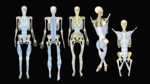
The term kinetic chain comes from engineering. Picture a series of rigid links connected by movable joints. Move one link and the others have to respond.
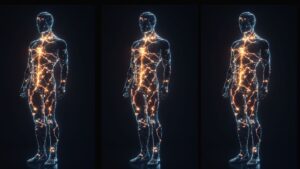
The body works the same way, but with an important difference. Our bones form the rigid links, and those links are connected by joints. Now add that joints are surrounded and crossed by muscles, tendons, fascia, and ligaments that guide movement, transmit force, and contribute to stability. Because these tissues connect one region to another, a restriction in a joint or increased tension in the surrounding soft tissues can change how movement and load are distributed throughout the entire chain.
And the body is a master compensator.
The lower-extremity chain begins at the floor and travels upward through the foot, ankle, tibia and fibula, knee, femur, hip, and pelvis. From there, it continues through the lumbar spine, thoracic spine, and ribcage, cervical spine, and head.
This is why I tell therapists not to chase pain. The place that hurts may simply be the place that is paying the bill for a restriction somewhere else.

In single-leg balance, both kinetic-chain conditions are at work: the lifted leg moves in an open chain, while the planted foot creates a closed chain that links motion at the ankle with the knee, hip, and pelvis.
Open Chain and Closed Chain
To understand the ankle, we first need to distinguish between open-chain and closed-chain movement. Lift your foot off the ground and move it around. That is open-chain movement. The foot is free, so the joints can move with relatively little influence from the floor.
Now plant the foot and bear weight through it. Everything changes. The foot is fixed against the ground, so movement has to travel upward through the leg. This is a closed chain. Almost everything that matters to your clients like standing, walking, climbing stairs, rising from a chair, or squatting, happens in a closed chain.
The defining property of a closed chain is that you cannot move one link in isolation. Pronate the foot and the tibia above it is obligated to rotate inward. Rotate the femur and the patella’s track in its groove changes whether you intended it to or not. The links are committed to one another.
The ankle is where this closed-chain relationship begins. Each time the foot accepts weight, the ankle must absorb load, adapt to the surface, and help direct force into the leg. How well it performs those jobs depends largely on two joints, the talocrural joint and the subtalar joint.
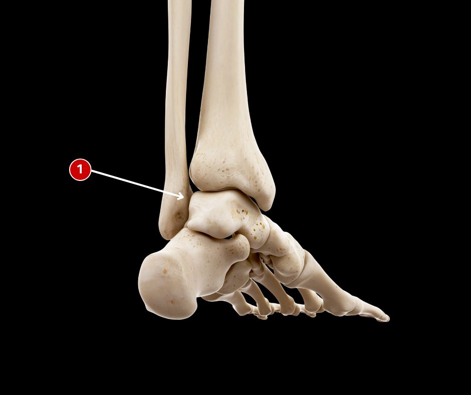
The talocrural joint is formed by the tibia, fibular, and talas.
The Talocrural Joint
The talocrural joint is the true ankle joint. It is a synovial hinge formed by the tibia, fibula, and talus, and its primary movements are dorsiflexion and plantar flexion. But the ankle behaves differently depending on whether the foot is free or planted.
Repeat the activity we performed previously but lift the foot toward the knee while the leg is off the ground. That is open-chain dorsiflexion. The foot moves toward the tibia.
Now place the foot on the floor and move the body forward. Because the foot cannot lift, the tibia must travel forward over the talus. That is closed-chain dorsiflexion, and it is essential for normal walking, squatting, stair climbing, and many other activities.
What happens when the ankle cannot dorsiflex fully? The body doesn’t simply stop moving. It compensates. The foot may collapse into additional pronation to manufacture more range. The heel may lift too early, reducing the efficiency of push-off. The knee may rotate or drift medially. The hip and pelvis may alter their movement to keep the body advancing.
The ankle loses motion, and the rest of the chain picks up the slack.
This is why limited dorsiflexion is one of the first things I look for when a client points to a painful knee. The knee may be where the client feels the problem, but the ankle may be where the movement problem begins.
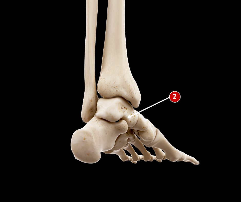
The subtalar joint is formed by the talus and calcaneus.
The Subtalar Joint
Just below the talocrural joint sits the subtalar joint, formed by the talus and calcaneus. The subtalar joint contributes to inversion and eversion and plays a central role in the larger triplanar movements of pronation and supination. It allows the rearfoot to adapt as we walk across uneven ground and helps the lower extremity respond to changing loads.
My mentor, osteopath Philip Greenman, called it the most important joint you did not know you had. Why did Greenman think this joint is so important? Because, where the talus goes, the tibia must follow.
The talus is unusual. It has extensive ligamentous connections to the tibia, fibula, calcaneus, and navicular, but no muscles attach directly to it. It cannot actively move itself. Instead, it responds to forces coming from the foot below and the leg above.
As the calcaneus moves, the talus changes position with it. Because the talus sits inside the mortise formed by the tibia and fibula, its movement influences rotation of the lower leg. When the rearfoot rolls inward, the talus adducts and plantar flexes as the tibia rotates inward. When the rearfoot moves outward, the talus and tibia move in the opposite direction.
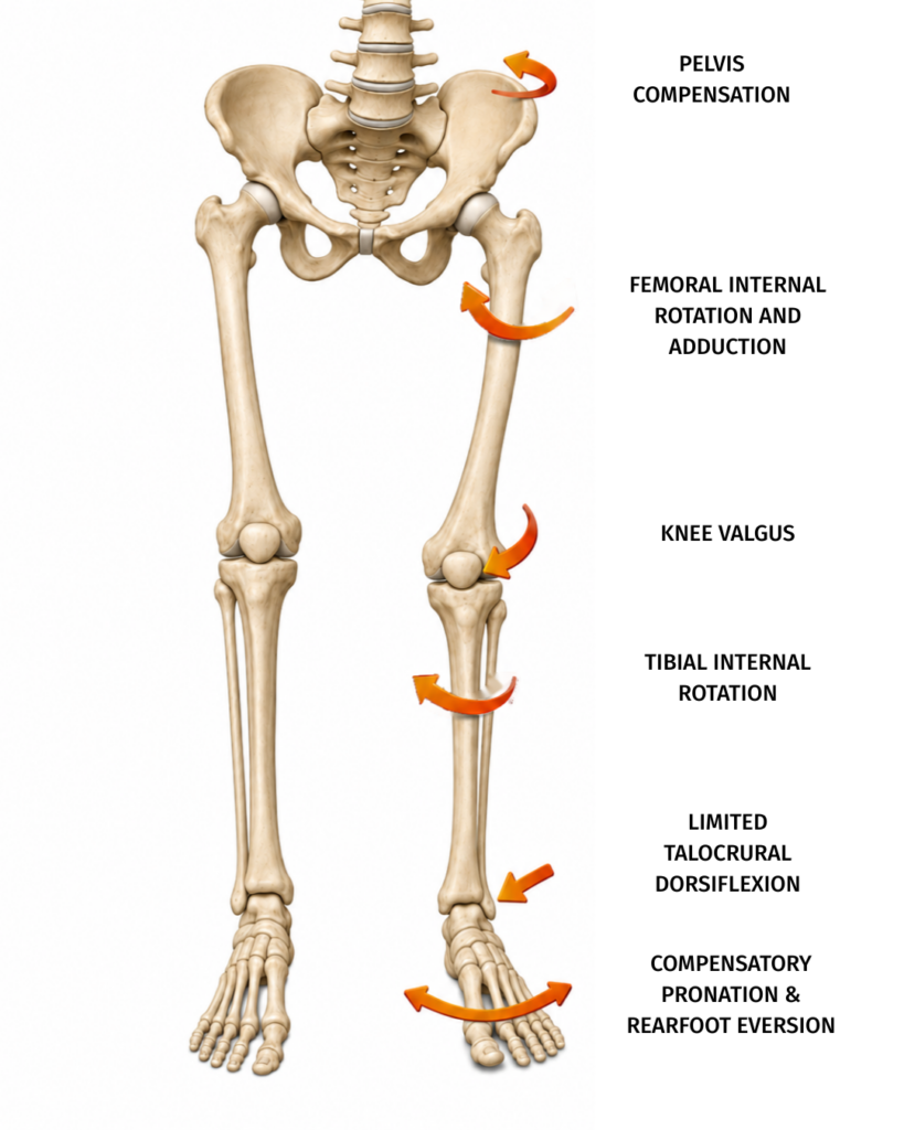
This illustration shows one possible closed-chain compensation pattern created by limited talocrural dorsiflexion. When the tibia cannot advance normally over the planted foot, the body may gain motion through increased pronation and rearfoot eversion at the subtalar joint. The talus and tibia rotate inward, and higher in the chain the femur may internally rotate and adduct. Together these contribute to dynamic knee valgus and compensatory movement at the pelvis. A restriction at the ankle can therefore change how the entire lower-extremity chain manages movement and load.
Every roll of the foot becomes a turn of the leg. This makes the subtalar joint an important mechanical converter. It helps transform movement between the relatively horizontal surfaces of the foot and the vertical orientation of the leg. Forces entering through the foot are translated into rotation that continues upward through the tibia, knee, and hip.
When the subtalar joint adapts well, those forces can be distributed throughout the chain. When it becomes stiff, compressed, or guarded, the body must find motion somewhere else. Once again, the knee or hip may be asked to produce movement the foot and ankle failed to provide. That’s how a restriction below becomes pain above.
Watch the Ankle and Foot Move
Watch the foot and ankle move through dorsiflexion, plantar flexion, inversion, and eversion courtesy of anatomy.app. Once you see these movements working together, it becomes easier to understand how losing even one of them can alter gait, reduce shock absorption, and create compensations throughout the kinetic chain.
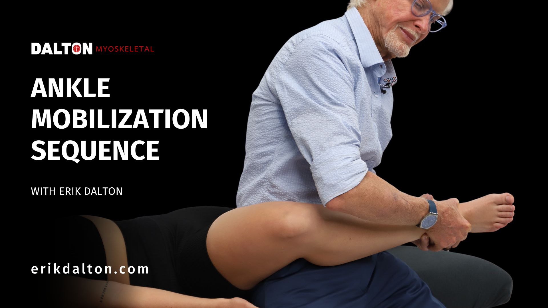
In the video at the top of the page, I demonstrate a supine ankle mobilization series you can easily add to a massage session. You will see how good body mechanics and mechanical advantage allow you to distract and decompress the ankle without muscling through the technique.
We will rock the joint, explore dorsiflexion and plantar flexion under distraction, and use gentle resisted movement when dorsiflexion feels restricted. We will also stabilize the calcaneus and introduce decompression and torsion at the subtalar joint to encourage more adaptable rearfoot motion.
The talocrural and subtalar joints form a critical bridge between the foot and the rest of the lower extremity. Together, they help the body move over the planted foot, adapt to the ground, and direct force into the leg. When either joint loses motion, the knee, hip, and pelvis may have to compensate.
So when a client complains of knee, hip, or even low-back pain, remember to look down the chain. Sometimes the best place to begin changing the pattern is at the ankle.