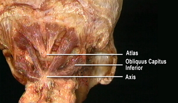
The second cervical vertebra, the axis, is considered the most important of all the neck’s bony structures partly due to its unique dural membrane attachment and also because of the powerful myofascial structures anchoring it from above and below (Image 1.). Deep suboccipital muscles that bind C2 to the occiput and atlas work in harmony with other muscles to balance the head on the neck.
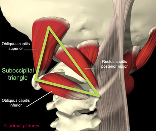
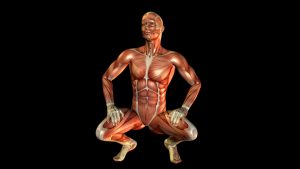
Several of the nerves that exit the upper cervical complex travel back over the top of the head to the forehead. These nerves must pass through a confined space called the suboccipital triangle (Image 2.). When the suboccipitals become irritated from physical strain, stomach sleeping or emotional stressors, they tighten…sometimes squashing the nerves that traverse the triangle.
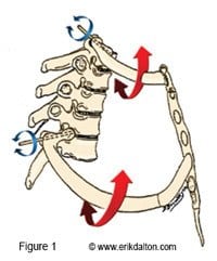
The obliquus capitis inferior (OCI) may be the most under appreciated of all the suboccipital muscles. Arising from the spinous process of C2 (axis) and inserting on the transverse process of C1 (atlas), their primary function is head-on-neck rotation (Image 3.). Notice in the image how the hypercontracted right OCI muscle is causing reciprocal inhibition and overstretching in the left…very common presentation in our chronic stomach sleeper population.
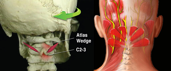
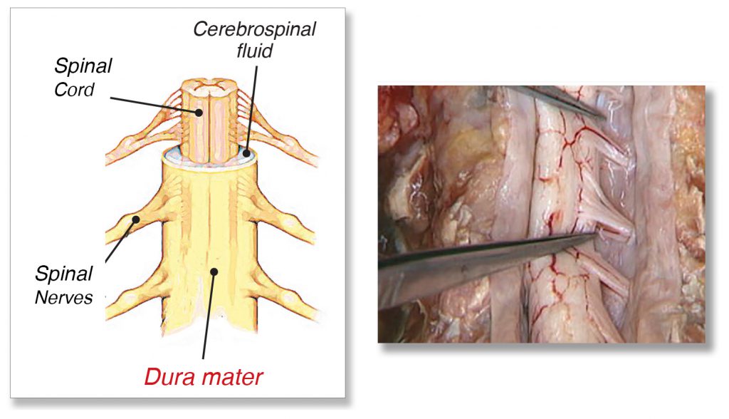
Visualize how a tight/short OCI on the right could restrain the atlas (and head) from left rotating. Yet, the person will often forget her stiff neck and quickly turn to look over her left shoulder. Can you see how this causes the right C2 facet joint to get crammed closed on C3 (Image 3.). This is a common area of dural membrane distortion as well as a key area of nerve impingement leading to headaches (Image 4.). Try the maneuver with a plastic spine.
When a rotated axis combines forces with an already overstretched dural tube from cranial or sacral distortions, a full-blown central nervous system assault may transpire. To relieve the client’s agonizing symptoms and restore healthy functioning, we must first have a clear picture of how the axis becomes misaligned and which techniques work best for releasing the disgruntled dural membrane.
Long-standing pain often fades in memory as dural techniques are properly applied through training programs devoted to this intriguing area of neuromechanics. Hands-on approaches for treating these conditions can best be learned by attending courses devoted exclusively to this very timely and rewarding body of work. By developing a comprehensive understanding of muscle/joint biomechanics involved in ‘dural drag’ and accompanying neck, head and low back pain, therapists can turn a therapeutic challenge into triumph.
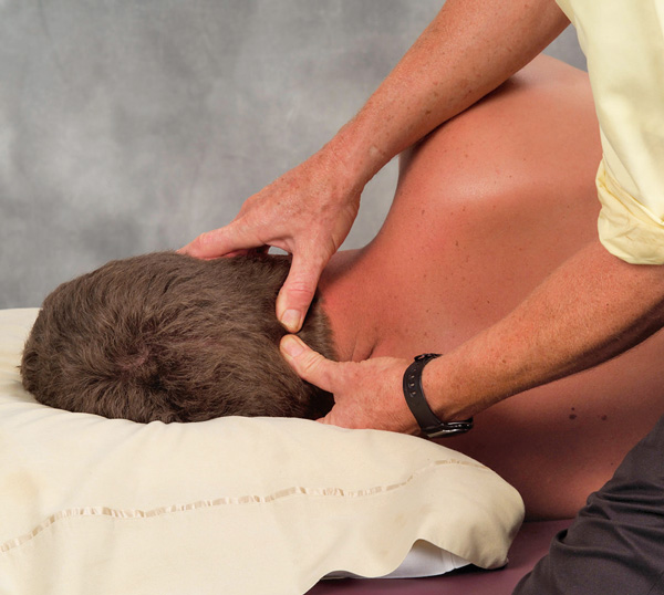
Creating Space in the Suboccipital Triangle
GOAL: Balance occiput on cervical column to relieve nerve pressure in suboccipital triangle
LANDMARK: Suboccipital ridge and spinous process of C2.
A. ACTION: Suboccipital Release Using Enhancers.
- Therapist’s right thumb locates spinous process of C2 and slides superiorly and slightly laterally to base of skull to contact rectus capitis posterior major.
- Therapist’s left thumb is placed directly below right thumb to contact rectus capitis posterior minor.
- Therapist drapes hands around client’s head so that thumb pads point towards one another.
- Client is instructed to gently cock head back against therapist’s thumbs to a count of five and relax.
- Therapist resists client’s extension efforts and holds for a mechanoreceptor release. Recall that the suboccipitals have no tendinous attachments and no GTOs at the occipital ridge.
On sale this week only!
Save 25% off the "Dalton Technique Treasures" eCourse
The “Dalton Technique Treasures” eLearning course is a compilation of some of Erik’s favorite Myoskeletal Alignment Techniques (MAT). Learn MAT techniques to assess and address specific sports injuries, structural misalignment, nervous system overload, and overuse conditions. ON SALE UNTIL July 29th! Get Lifetime Access: As in all our eLearning courses, you get easy access to the course online and there is no expiry date.

