Treating Kinetic Chain Kinks
Recent manual and movement therapy blogs tout the importance of thoracic spine (t-spine) mobility as if it were a new discovery. But is it? Structural integration innovator Ida Rolf determined that her work could be enhanced by first freeing the rib cage, chest wall, and diaphragm. Similarly, another great educator, Philip Greenman, DO, dedicated two chapters in his textbook “Principles of Manual Medicine” to assessment and treatment of t-spine and rib cage mobility issues. So why is this suddenly a hot topic? Simply put: kinetic chain awareness.

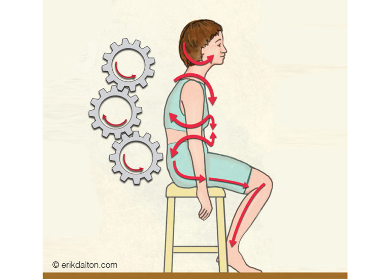
For instance, during gait evaluation, it’s easy to visualize how lack of ankle mobility may affect knee function, or how an adhesive hip capsule could cause pelvic bowl compensations that destabilize a sacroiliac joint or the low back. However, confusion often arises when one observes the t-spine and rib cage. Although the t-spine has twice the rotational capacity of the lumbar spine, it is sometimes hard to imagine this sturdy-looking structure being very flexible.
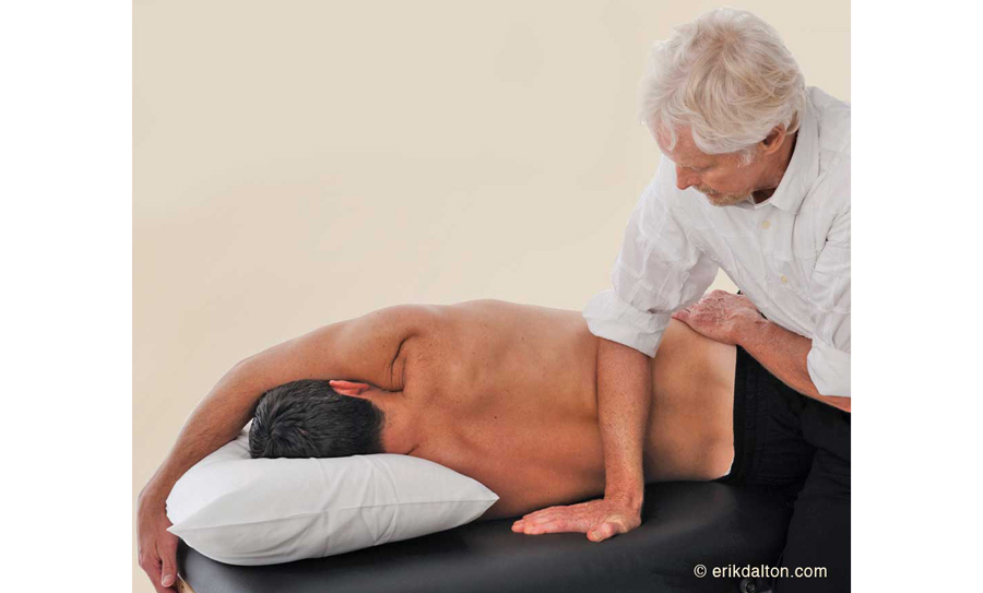
T-spine hypomobility has become so commonly accepted in our society that people rarely notice they have a problem. Nearly everyone slumps when sitting, and few perform the types of exercises that require a full range of spinal motion. Those who spend hours at computers sacrifice t-spine mobility for stability, as joint and ligament proprioceptors designed to inform the brain where it is in space become lazy. Conversation between body and brain grows difficult and unreliable. Eventually, coordination, balance, and movement become limited and painful.
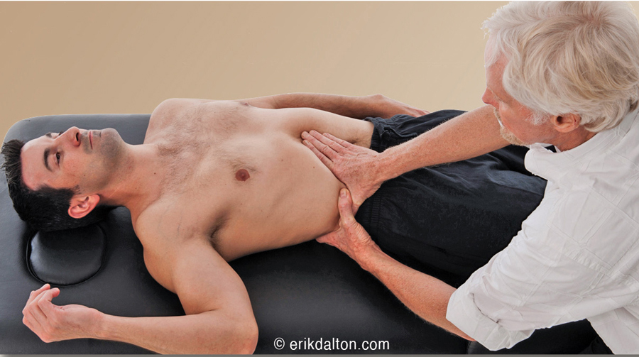
When you shoot a rubber band, it will be propelled a greater distance the farther back it is pulled. Similarly, the greater your myoskeletal mobility, the greater your range of motion, and the more tension (and therefore power) you’ll be able to generate. This particularly applies to competitive athletes. Strength without the ability to move freely is pointless. Any compound movement requiring precision and communication between connective tissue, joints, and the brain will be more difficult, and the risk of injury—or reinjury—that much higher. Power, output, and speed are all compromised by reduced joint mobility.
References
1. Kamkar A, Cardi-Laurent C, Whitney SL. Conservative management of superior subluxation of the first rib. J Sport Rehabil. 1992;1(4):300–316.
2. DeStefano L. Greenman’s Principles of Manual Medicine. Philadelphia, PA: Lippincott Williams & Wilkins; 2011.
On sale this week only!
Save 25% off the Upper Body Course!
NEW! Enhanced video USB format!!!
Learn unique approaches to work with the shoulder girdle, arms, neck, and torso, this course will prepare you to relieve painful myoskeletal issues in the upper body. Through video demos and animation, you’ll learn to identify several compensatory movement patterns and their associated reflexogenic pain. With this understanding of where the true source of problems arise, you’ll be able to develop highly effective treatment protocols and deliver lasting results. This course provides you with the skills you need to confidently relieve pain issues in the upper body. (20 CE)
Bonus: When you order the Home Study version, get the eLearning course and and an additional 2CE Ethics eCourse for free!
Save 25% off the Upper Body Course this week only. Offer expires Sunday, March 1st. Click the button below for more information and to purchase the course for CE hours and a certificate of completion to display in your office.

