fbpx

Addressing SI Joint Syndrome

From Dynamic Lower Body course

Myoskeletal technique for hip and back pain

In the early 20th century, sacroiliac joint syndrome (SIJ) was the most common medical diagnosis for low back pain, which resulted in that period being labeled the “Era of the SI Joint.” Any pain emanating from the low back, buttock or adjacent leg usually was branded and treated as SIJ. However, this medical mindset came to a screeching halt in 1934, when Jason Mixter, MD, published an article on the intervertebral disc in The New England Journal of Medicine.1His landmark report challenged the popular understanding of sciatica and helped establish surgery’s prominent role in the management of sciatica at the time. Throughout the years, discectomy surgery has increased in popularity, causing many to define this period as “Dynasty of the Disc.”
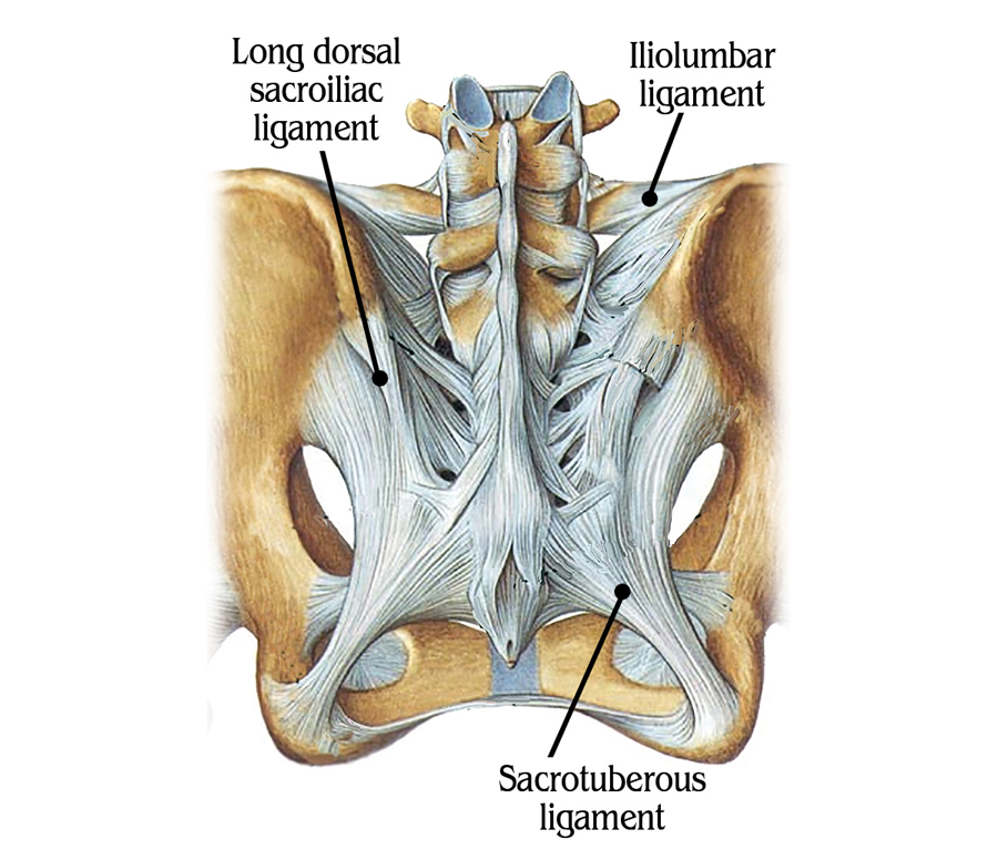
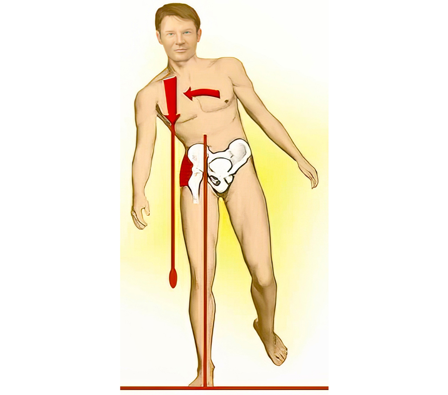
Image 1. Pelvic Ligaments
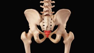
SI joint syndrome continued its fall from fashion due to the lack of reliable clinical studies confirming its very existence. Although many manual therapists quietly continued treating this disorder with some success, no one was able to put forward a convincing biomechanical theory explaining how the sacrum becomes stuck “crooked” between the two innominate bones. Physicians were reluctant to envision a joint with so little movement causing so much pain, while manual therapists countered that its limited motion was vital to proper lumbar spine functioning. Since most SI joints only move about 2 to 4 millimeters during weight bearing and forward bending, they are described as a gliding-type joint. This motion is quite different from the hinge-type articulation at the knee or the ball-and-socket motion of the hip. Considered a viscoelastic joint, the SI’s major movement comes from ligamentous stretching (Image 1.). Therefore, its primary function within the pelvic girdle is to provide shock absorption for the spine by stretching in various directions. When sacroiliac joints work in perfect harmony with the third bony articulation of the pelvis (symphysis pubis), a marvelous self-locking mechanism develops that helps us walk. Aided by power generated by the hip abductors (gluteus medius/ minimus, TFL and piriformis), the pelvic joints brace the weight-bearing side during gait. This locking system, termed force closure, allows smooth transference of body mass from one leg to the other (Image 2.).
Image 2. Hip abductors (gluteus medius/ minimus, TFL and piriformis) force-close the ipsilateral SI joint during the stance phase
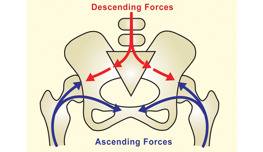
Image 3. Pelvis is designed to absorb ascending and descending forces
Although no muscles directly bind down the three pelvic joints, when working synchronously with the SI ligaments they provide the pelvis − “the great adapter” – with a remarkable antigravity spring system that can absorb both ascending and descending forces (Image 3.). Sacroiliac pain tends to be low down; often more to one side, and may extend into the buttock, back of the thigh, and occasionally the calf. It can start suddenly, but in chronic cases, strained joint and ligament mechanoreceptors trigger protective muscle spasm that cause dull, nagging buttock pain or pins and needles down the posterior thigh. During the aging process, there is decreased motion at the sacroiliac joint due to normal wear and tear and accompanying calcification. This is a perfect example of how the body’s innate wisdom will sacrifice “complexity of motion for stability.” An interesting note is that the age with highest incidence of disabling back pain (25-45 years) is the same age at which the greatest amount of motion is available in the sacroiliac joints. It’s not uncommon for SI joints to stiffen and permanently lock with age. So, it behooves massage therapists to begin incorporating specialized soft-tissue SI joint mobilization maneuvers to help calm the nervous system, relieve protective guarding, and encourage stability and more functional movement through the pelvis. Image 4.
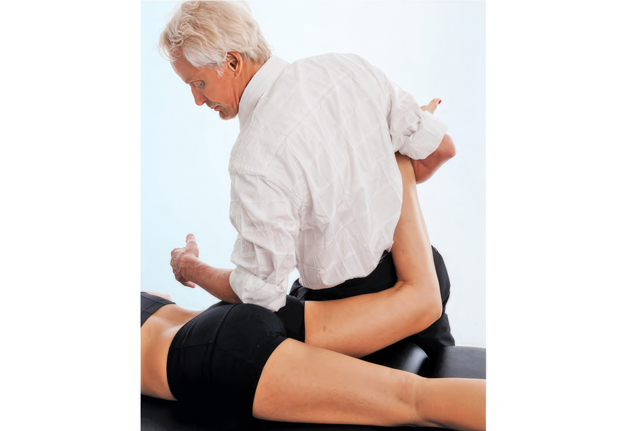
Image 4. A counterforce occurs as the therapist’s left forearm hooks the client’s hip external rotators while her right leg is taken into internal rotation. Therapist assesses and releases hip hypertonicity.
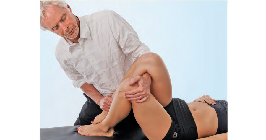
Ligamentous laxity can also be a problem. Traumatic blows to the hip may result in SI joint ligament hypermobility, and over-stimulation of the joint’s mechanoreceptors. As the articulating joint surfaces become jarred loose, ligament microtearing may create an inflammatory response. As sensitized chemoreceptors and mechanoreceptors bombard the spinal cord and brain with noxious stimuli, the brain may choose to layer the area with protective muscle guarding to prevent further insult. This is the beginning of a therapeutically challenging pain/spasm/pain cycle that often is hard to break. It is possible, however, to down-regulate the pain emanating from hypermobile joints by first restoring proper pelvic alignment using techniques such as the one shown in Image 5., and then addressing core strength issues via home retraining exercises.
Image 5. To posteriorly rotate client’s right ilium, the therapist snakes his left arm under her right leg and grasps her left knee. Therapist left rotates client’s hips and applies a counterforce between both hands to decompress the right fixated SI joint.

Femoroacetabular, SI and Lumbar Spinal Joints

Although the three bones of the pelvis frequently are at the seat of a “primary” lesion, I’ve found that a missing key in successful correction of SI pain is mobilizing motion-restricted femoroacetabular (hip) joints. For the pelvis to effectively absorb the forces imposed upon it, the hips must be aligned and functioning properly. Normally, it’s not the gross motions creating dysfunction within the hip’s truncated joint capsule, but restrictions of minor movements caused by such things as iliofemoral ligament adhesions (Image 6.). Therefore, a rational treatment approach would begin with mobilization of the adhesive anterior hip capsule as shown in Image 7., followed by the iliosacral and sacroiliac alignment techniques presented above.
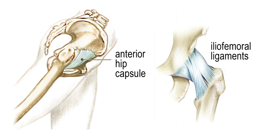
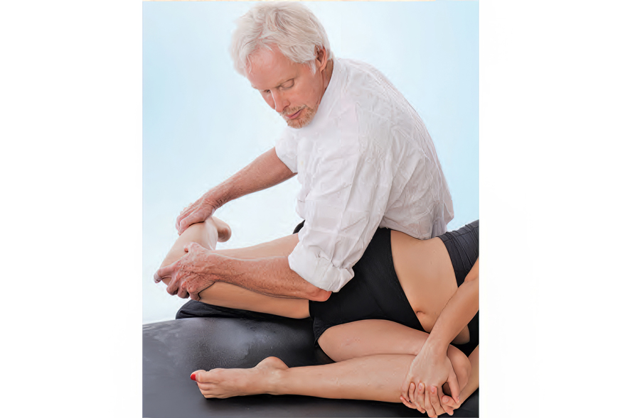
Image 6. The Iliofemoral ligament can bind down the anterior hip capsule preventing ipsilateral hip extension during gait.
Image 7. Therapist uses a graded exposure stretch to release an adhesive hip capsule and rectus femoris. Retest for improved hip extension during gait.

Legendary neurologist and physiotherapist Vladimir Janda, MD, reminds us that: “Any alteration in joint function caused by capsular restriction or loss of joint play affect muscles that cross the dysfunctional joint either through inhibition (weakening) or facilitation (tightening).” Many in today’s flexion-addicted society suffer from anterior hip capsule adhesions and tight iliopsoas muscles that bind the right femur into a flexed position, preventing adequate hip extension during gait. Yet we have to walk … so what happens biomechanically? As the right leg swings back into extension, the neurologically shortened iliopsoas and fibrosed right hip capsule prevent full hip extension causing the pelvic bowl to anteriorly rotate. This results in facet joint and disc compression, overstretching of pelvic ligaments, and compensatory lumbar scoliosis.

Summary

A stable pelvis is achieved through proper upper and lower quadrant balance and is essential for long-term low back and SI joint health. In the presence of pelvic instability, the brain will attempt to stabilize the lumbosacral joint by layering the area with protective spasm, but it does little good for therapists to try and release the muscle hypertonicity until the hip and pelvic alignment problems are corrected. Myoskeletal techniques that include low-force mobilization and graded-exposure stretching help the brain recognize and reorganize neural input resulting in less reactive muscle spasm, less pain and happier clients.

References

  1. Parisien RC, Ball PA. William Jason Mixter (1880-1958). Ushering in the “dynasty of the disc.” Spine 1998;23(21):2363-6.
  2. Janda V. Treatment of chronic back pain. Journal of Manual Medicine1992;6:166-8.

On sale this week only!

Save 25% off the Art of MAT course!

NEW! Enhanced video USB format

Take an inside look at Myoskeletal Alignment Techniques in action. Follow along with Erik Dalton as he uses MAT techniques to treat real people with real problems. Click the button below for more information and to purchase the course for CE hours and a certificate of completion to display in your office. BONUS: When you purchase the home study course, receive the eCourse for free!

Save 25% this week only. Offer expires November 25th!

Free subscription to the Technique Tuesday Newsletter

Receive an in-depth article like the one on this page along with a technique video every week in your inbox with no subscription fees.